Method for detecting immune body affinity

A technology for detecting antibodies and affinity, which is applied in the field of medicine and biology, can solve the problems of long time required, achieve good repeatability, improve detection efficiency, and high sensitivity

Inactive Publication Date: 2008-05-07
SUREXAM BIO TECH
View PDF0 Cites 3 Cited by
  • Summary
  • Abstract
  • Description
  • Claims
  • Application Information

AI Technical Summary

Problems solved by technology

At the same time, the antigen coating needs to be overnight at 4°C, and there are multiple washing steps, which take a long time

Method used


Image

  • Method for detecting immune body affinity

Examples


Embodiment 1

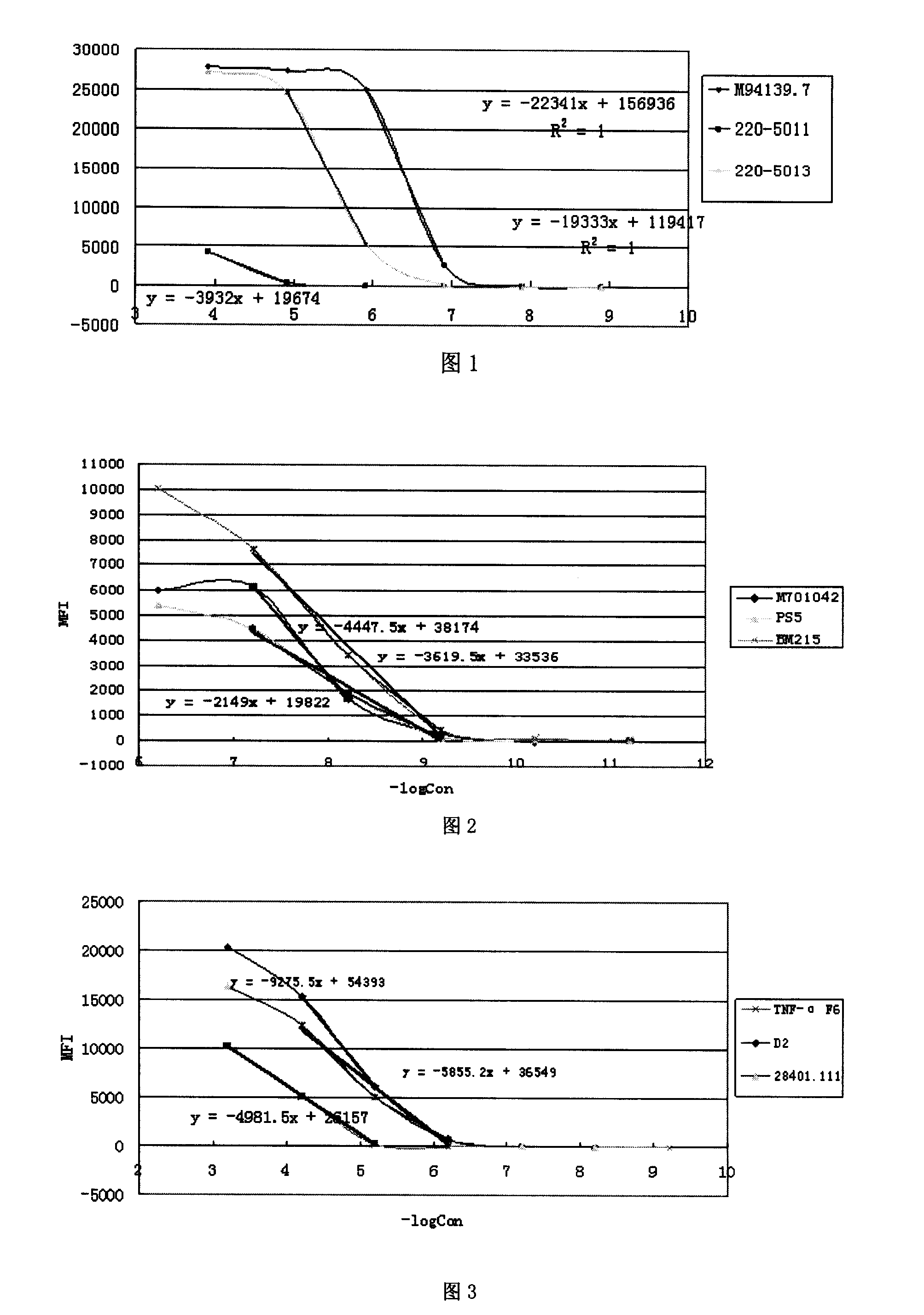
[0064] Take the affinity detection of three different antibodies (cloning numbers M94139.7, 220-5011, and 220-5013) to β-HCG (β-subunit human chorionic gonadotropin) as an example.

[0065] The steps of the antibody affinity detection method are as follows:

[0066] Antigen-coated microspheres:

[0067] -Use a vortex oscillator or ultrasonic to suspend the microspheres (Luminex, USA), about 20s;

[0068] - Take 50μL of microspheres in a 1.5ml centrifuge tube and centrifuge at a speed of ≥8,000g for 1-2min;

[0069] -Remove the supernatant, resuspend the microspheres in 100 μL double distilled water, suspend the microspheres with a vortex shaker or ultrasonic wave for about 20 seconds, and centrifuge at a speed of ≥8000g for 1-2 minutes;

[0070] - Discard the supernatant, add 80 μL of phosphate buffer (pH 6.2), vortex for about 20 seconds, and ultrasonically suspend the microspheres for about 20 seconds;

[0071] -Add 10μL 50mg / mL Sulfo-NHS and mix gently with a vortex shak...

Embodiment 2

[0112] Affinity detection of three antibodies (cloning numbers: M701042, PS5, BM215) of PSA (Prostate Specific Antigen).

[0113] The method used is the same as that of Example 1. The result is as follows,

[0114] Table 2

[0115] μg / ml

mol / L

negative logarithm of concentration

M701042 (MFI value)

PS5 (MFI value)

BM215 (MFI value)

100

10

1

0.1

0.01

0.001

6.25E-07

6.25E-08

6.25E-09

6.25E-10

6.25E-11

6.25E-12

6.20412

7.20412

8.20412

9.20412

10.20412

11.20412

6323.5

6479

2031.5

640

336.5

438.5

5369

4481.5

1910.5

183.5

97

52.5

10054.5

7657

3447.5

418

125

57

[0116] The result is calculated as follows:

[0117] Calculation of the affinity constant of the M701042 antibody:

[0118] y=(...

Embodiment 3

[0122] Affinity detection of three antibodies to TNF-α (Tumor necrosis factor-alpha, tumor necrosis factor α) (cloning numbers are respectively TNF-αF6, D2, 28401.111).

[0123] The method used is the same as that of Example 1. The result is as follows,

[0124] table 3

[0125] μg / ml

mol / L

negative logarithm of concentration

TNF-αF6 (MFI value)

D2 (MFI value)

28401.111 (MFI value)

100

10

1

0.1

0.01

0.001

0.0001

0.000625

6.25E-05

6.25E-06

6 25E-07

6.25E 08

6.25E-09

6.25E-10

3.204119983

4.204119983

5.204119983

6.204119983

7.204119983

8.204119983

9.204119983

10233.5

5137.5

270.5

0

0

2

16

20352

15397.5

6122

779

70.5

15.5

16397

12433

5077

722.5

73

16

[0126] The res...


PUM


Abstract

The invention discloses a method for detecting antibody affinity. The detection method mainly includes the steps of (1) antigen-coated microspheres, (2) detection of antibody affinity and (3) data processing. The method for detecting antibody affinity has the advantages of simplicity, quickness, high sensitivity, good repeatability, more stable and reliable detection results, and the like.

Description

technical field [0001] The invention belongs to the category of medical biology, and in particular relates to a method for detecting antibody affinity. technical background [0002] Antibody affinity is one of the important parameters to determine the properties of antibodies, and at the same time, it is also an important indicator of the stability of monoclonal antibodies (McAb) secreted by hybridomas. It indicates how tightly the antibody binds to the antigen. Antibodies with high affinity are useful for quantitative detection and oriented diagnosis and treatment, while antibodies with low affinity are better for immunopurification. Therefore, in order to select an antibody with a certain affinity according to the purpose of the application, it is necessary to test the affinity of the antibody first. [0003] Antibody affinity refers to the firmness of antibody-antigen binding, which is often expressed by the affinity constant Ka. Ka is the ratio of the forward / reverse ...

Claims


Application Information

Patent Timeline
Patent Type & Authority Applications(China)
IPC IPC(8): G01N33/543G01N33/531
Inventor 彭臻菲杨惠夷许嘉森
Owner SUREXAM BIO TECH
Why Eureka
Try Eureka