Liver subsection method based on CT image and system thereof

A technology of liver segmentation and liver, which is applied in the field of liver segmentation method and its system, can solve the problems of inability to quickly and conveniently correct the grading results, segmentation errors, and inability to perform interactive operations, etc., to achieve easy modification, convenient grading, and improved results Effect

Inactive Publication Date: 2012-03-28
HUAZHONG UNIV OF SCI & TECH
View PDF1 Cites 1 Cited by
  • Summary
  • Abstract
  • Description
  • Claims
  • Application Information

AI Technical Summary

Problems solved by technology

If the local specificity of blood vessels may cause grading errors, this method cannot be operated interactively, so it is not convenient to quickly correct the grading results and cause segmentation errors

Method used


Image

  • Liver subsection method based on CT image and system thereof
  • Liver subsection method based on CT image and system thereof
  • Liver subsection method based on CT image and system thereof

Examples


Embodiment Construction

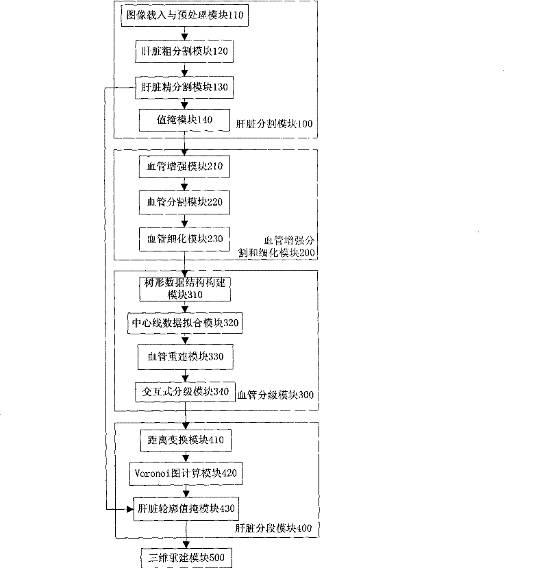
[0048] The invention is a complete liver segmentation method for MSCTP sequence images. The liver segmentation of the present invention is divided according to the areas to which different branches of the portal vein belong, and the liver segmentation mainly includes five parts: liver segmentation, blood vessel segmentation and thinning, blood vessel classification, liver segmentation and three-dimensional reconstruction.

[0049] The technical solutions of the present invention will be further described in detail below in conjunction with the accompanying drawings and examples.

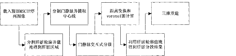
[0050] Such as figure 1 Shown, the inventive method comprises the following steps:

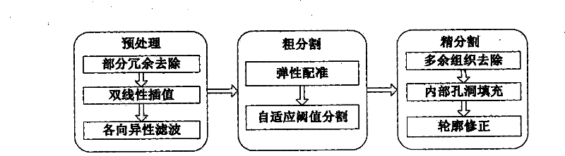
[0051] (1) Load the portal venous phase and arterial phase sequence images of abdominal MSCTP, and automatically segment the liver. The flow chart is as follows image 3 As shown, the specific steps are as follows:

[0052] (1.1) Preprocessing. The sequence images of the portal venous phase and the arterial phase...


PUM


Abstract

The invention discloses a liver division method based on a CT image, which comprises the following steps: firstly, an MSCTP artery sequence image and a hepatic portal vein sequence image of the abdomen are pretreated, and the outline of the liver is automatically divided to obtain the liver image; secondly, a multi-scale filtering method based on a Hessian matrix is used for strengthening blood vessels, a dividing method such as region enlargement, and the like is used for dividing the hepatic portal vein, and a three-dimensional topology thinning method is used for picking up the central line of the hepatic portal vein; the blood vessels are identified mutually in classification; distance change and Voronoi arithmetic are then used for calculation, and the outline of the liver is used for covering values to obtain division results; the three-dimensional liver division results are finally reconstructed. The system comprises a liver division module, a blood vessel strengthening dividing and a thinning module, a blood vessel classifying module, a liver dividing module and a three-dimensional reconstructing module. The invention can reduce noise as well as avoid the vagueness of the boundary of the liver, effectively and exactly divides the outline of the liver, improves the division quality of the hepatic portal vein tree, and realizes accurate and rapid division of the liver.

Description

technical field [0001] The invention belongs to the field of medical image processing and application, and in particular relates to a liver segmentation method and a system thereof. The invention can effectively segment the liver, reconstruct a three-dimensional model of the liver, and can visually display the shape of each segment of the liver. Background technique [0002] The liver is the largest solid organ in the human body. Anatomical studies have shown that the liver is divided into eight liver segments, and each liver segment has an independent pipeline system, which can be used as a surgical resection unit. With the development of liver transplantation and liver resection in recent years, the segmental anatomy of the liver has attracted more and more attention from surgeons. Multi-slice spiral CT (multi-slice spiral CT, MSCT) is widely used in clinical diagnosis, and its enhanced scan images are divided into three-phase sequence images of arterial phase, portal ven...

Claims


Application Information

Patent Timeline
Patent Type & Authority Patents(China)
IPC IPC(8): G06T17/00G06T7/00A61B6/03
Inventor 张智万少朋郭睿丁亮刘晶晶胡道予谢庆国
Owner HUAZHONG UNIV OF SCI & TECH
Why Eureka
Try Eureka