
Laser speckle blood current imaging and analyzing method

A technology of laser speckle and analysis method, applied in the field of laser speckle blood flow imaging analysis, to achieve the effect of wide application range and high time resolution

Inactive Publication Date: 2009-07-22
HUAZHONG UNIV OF SCI & TECH
View PDF2 Cites 49 Cited by
  • Summary
  • Abstract
  • Description
  • Claims
  • Application Information

AI Technical Summary

Problems solved by technology

[0003] The purpose of the present invention is to provide a laser speckle blood flow imaging analysis method, which can solve the limitation that the existing laser speckle imaging method cannot take into account the spatial resolution and time resolution, and realize the two-dimensional analysis of blood flow in biological tissues. Real-time high-resolution imaging of distribution

Method used


Image

  • Laser speckle blood current imaging and analyzing method
  • Laser speckle blood current imaging and analyzing method
  • Laser speckle blood current imaging and analyzing method

Examples


Embodiment Construction

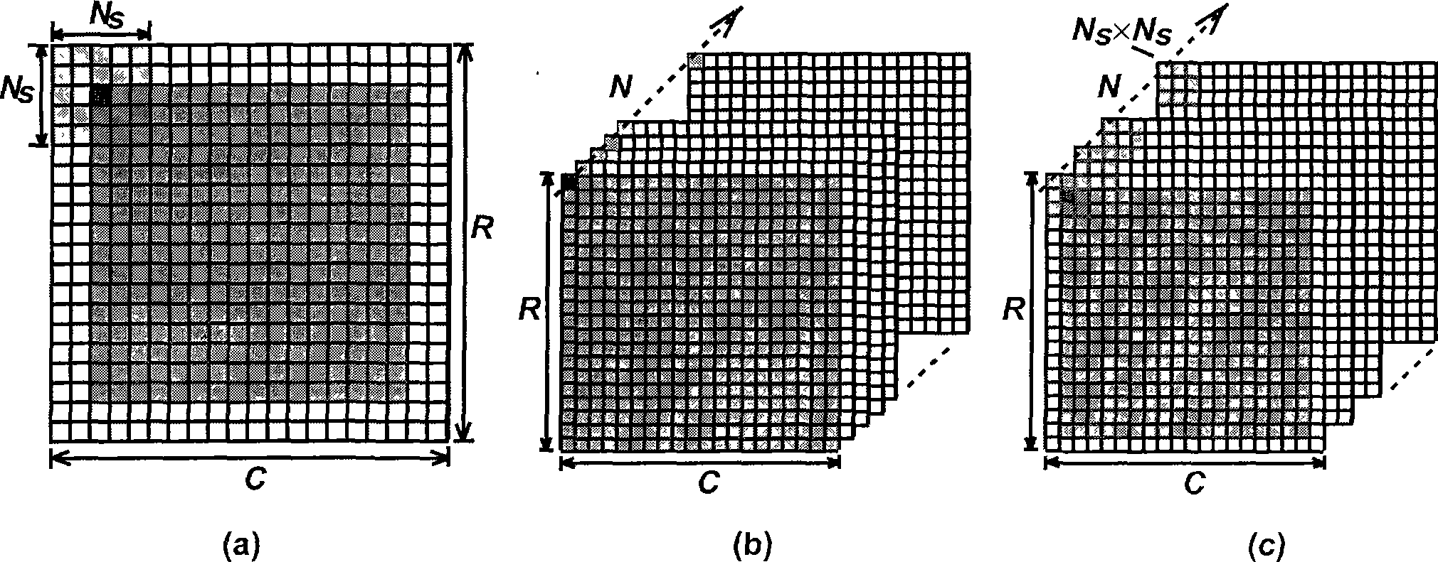
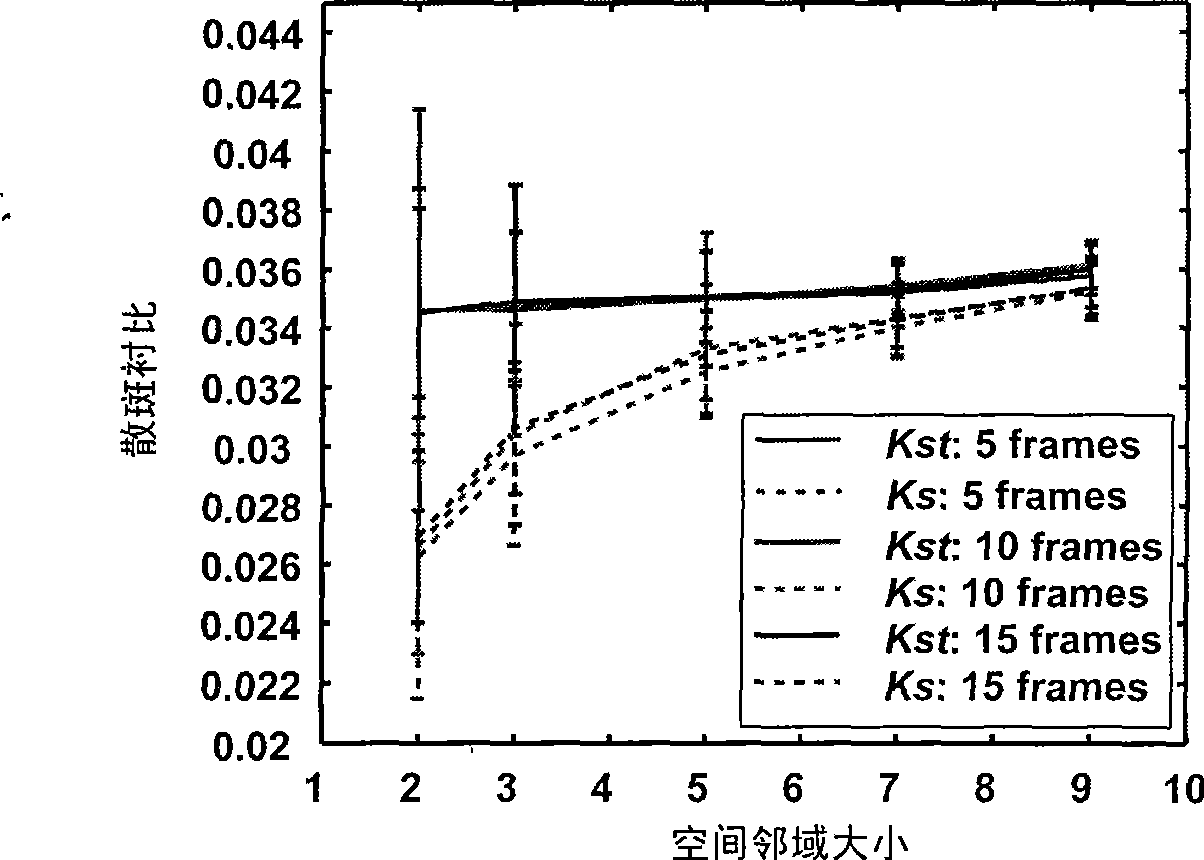
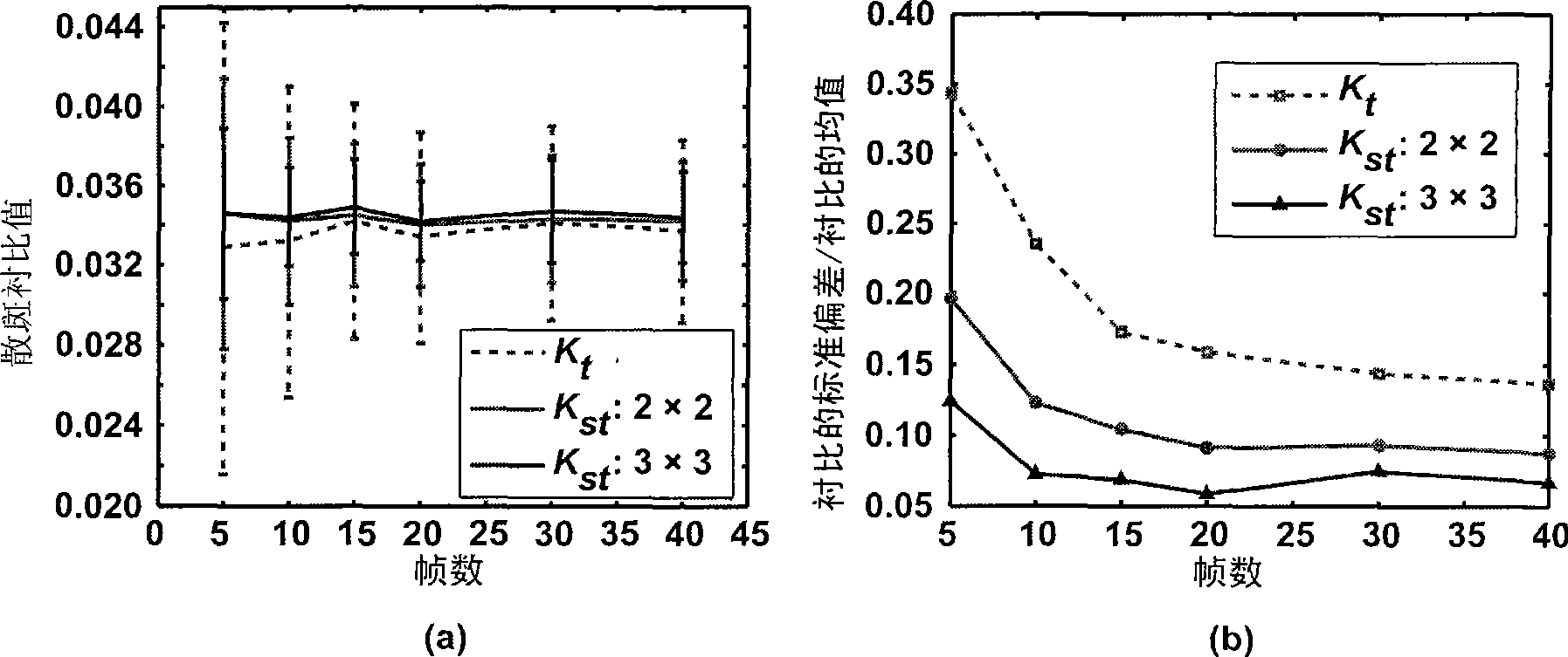
[0018] The reconstruction of blood flow distribution images in biological tissues needs to use several frames of laser speckle images collected in each spatial neighborhood where blood flow needs to be measured, and perform joint statistics in time domain and space domain on the collected laser speckle image sequences Characteristic analysis, calculate the statistic of light intensity of all pixels (i.e. image grayscale) in each spatial neighborhood corresponding to the laser speckle image, and use this statistic to reflect the blood flow velocity at the biological tissue corresponding to the pixel; By traversing all pixels in the image in this way, a high-resolution two-dimensional biological tissue blood flow distribution image can be obtained. as attached figure 1 As shown in (c), the spatial neighborhood used to calculate the contrast value is smaller than that used by the spatial contrast algorithm, and the number of time series image frames required is less than that req...


PUM


Abstract

The invention discloses a laser speckle blood stream imaging analysis method. The method comprises: irradiating a laser beam on an object to be detected and continuously acquiring N frames of laser speckle images reflected by the object to be detected according to identical exposure time and frame intervals; taking pixels from a Ns*Ns spatial neighborhood corresponding to identical positions in each frame of image to form a pixel set; calculating a contrast Kst within the spatial neighborhood and then calculating the blood stream speed V(i, j) of a corresponding biological tissue by means of a relational expression of the contrast and the blood stream speed; and taking the blood stream speed value corresponding to each pixel as grey scale to establish a two-dimensional blood stream speed distribution map. The laser speckle blood stream imaging analysis method is based on space-time combined analysis of laser speckle blood stream imaging to carry out high time and spatial resolution imaging on blood stream of a biological issue, thereby realizing real-time, dynamic and high time and spatial resolution monitoring of two-dimensional blood stream distribution, blood vessel form and blood stream dynamic changes of the biological tissue. The analysis method is suitable for researching brain function imaging, neurophysiology, disease pathology and drug effect evaluation.

Description

technical field [0001] The invention belongs to the field of biological tissue blood flow imaging methods, in particular to a laser speckle blood flow imaging analysis method, which is especially suitable for studying local blood flow distribution and vascular morphology of biological tissues under physiological and pathological conditions with high temporal and spatial resolution. . Background technique [0002] Laser speckle blood flow imaging is widely used to study local two-dimensional blood flow changes in biological tissues under physiological and disease states. However, the existing analysis methods of laser speckle blood flow imaging cannot take into account the time resolution and spatial resolution of the image at the same time. Such as the patent document "Using Laser Speckle Imager to Monitor the Time-Space Response Characteristics of Mesenteric Microcirculation Blood Flow" (its publication number is CN1391869, and its publication date is 2002.07.12), and its ...

Claims


Application Information

Patent Timeline
IPC IPC(8): A61B5/02A61B5/026
Inventor 李鹏程骆清铭邱建军
Owner HUAZHONG UNIV OF SCI & TECH
Learn More