Novel use of cyanine dye in detection of G-quadruplex DNA
A technology of cyanine dye and quadruplex is applied in the new application field of cyanine dye in the detection of G-quadruplex structure DNA, which can solve the problems of high price, difficult separation and purification, and high equipment requirements, and can overcome the long cycle and cost. low cost effect
- Summary
- Abstract
- Description
- Claims
- Application Information
AI Technical Summary
Problems solved by technology
Method used
Image
Examples
Embodiment 1
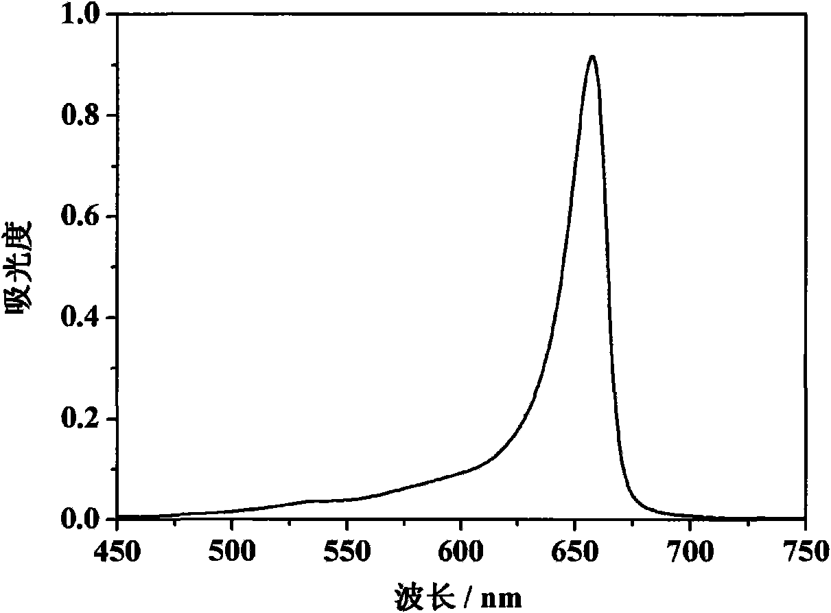
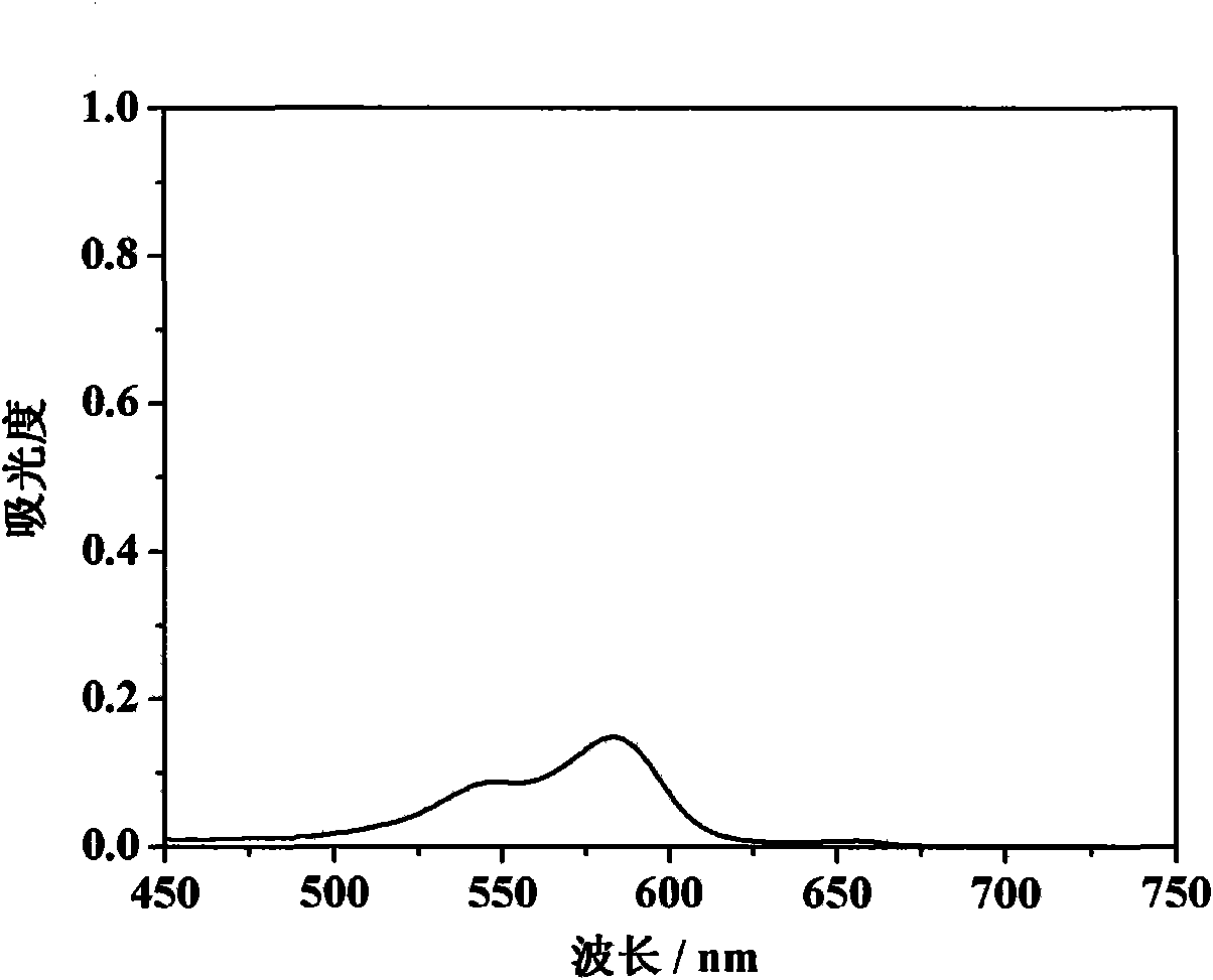
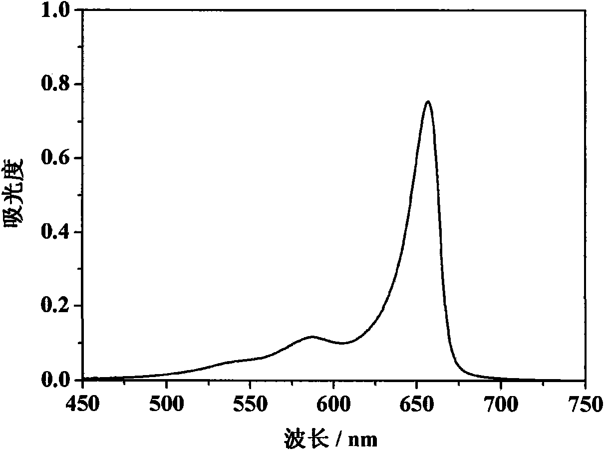
[0065] Embodiment 1, the detection of G-quadruplex structure DNA
[0066] 1. Preparation of reaction solution
[0067] 1) Solution A
[0068] Add 40 μL 200.0 μM methanol (excellent grade) solution of cyanine dye to a 2ml volumetric flask; add 60 μL 200 μM phosphate buffer solution (pH 6.0) of sample 1, dilute to volume with phosphate buffer solution (pH 6.0), mix well, Solution A is obtained. In solution A, the molar ratio of sample 1 DNA to cyanine dye is 1.5:1.
[0069] 2) Solution B
[0070] Add 40 μL 200.0 μM methanol (excellent grade) solution of cyanine dye to a 2ml volumetric flask; add 60 μL 200 μM phosphate buffer solution (pH 6.0) of sample 2, dilute to volume with phosphate buffer solution (pH 6.0), mix well, Solution B is obtained. In solution B, the molar ratio of sample 2 DNA to cyanine dye is 1.5:1.
[0071] 3) Solution C
[0072] Add 40 μL of 200.0 μM methanol (excellent grade) solution of cyanine dye to a 2 ml volumetric flask, dilute to volume with pho...
Embodiment 2
[0087] Embodiment 2, the detection of G-quadruplex structure DNA
[0088] 1. Preparation of reaction solution
[0089] 1) Solution A
[0090] Add 40 μL of 200.0 μM methanol solution of cyanine dye to a 2 ml volumetric flask; add 20 μL of 200 μM phosphate buffer solution (pH 6.0) of sample 1, dilute to volume with phosphate buffer solution (pH 6.0), and mix to obtain solution A. In solution A, the molar ratio of sample 1 DNA to cyanine dye is 0.5:1.
[0091] 2) Solution B
[0092] Add 40 μL of 200.0 μM methanol solution of cyanine dye to a 2 ml volumetric flask; add 20 μL of 200 μM phosphate buffer solution (pH 6.0) of sample 2, dilute to volume with phosphate buffer solution (pH 6.0), and mix to obtain solution B. In solution B, the molar ratio of sample 2 DNA to cyanine dye is 0.5:1.
[0093] 3) Solution C
[0094] Add 40 μL of a 200.0 μM methanol solution of cyanine dye molecules into a 2 ml volumetric flask, dilute to the volume with phosphate buffer (pH 6.0), and mix ...
Embodiment 3
[0106] Embodiment 3, the detection of G-quadruplex structure DNA
[0107] 1. Preparation of reaction solution
[0108] 1) Solution A
[0109] Add 40 μL of 200.0 μM pure methanol solution of cyanine dye to a 2ml volumetric flask; add 160 μL of 200 μM phosphate buffer solution (pH 8.0) of sample 1, dilute to volume with phosphate buffer solution (pH 6.0), and mix to obtain solution A. In solution A, the molar ratio of sample 1 DNA to cyanine dye is 4:1.
[0110] 2) Solution B
[0111] Add 40 μL of 200.0 μM pure methanol solution of cyanine dye to a 2 ml volumetric flask; add 160 μL of 200 μM phosphate buffer solution (pH 8.0) of sample 2, dilute to volume with phosphate buffer solution (pH 6.0), and mix to obtain solution B. In solution B, the molar ratio of sample 2 DNA to cyanine dye is 4:1.
[0112] 3) Solution C
[0113] Add 40 μL of 200.0 μM pure methanol solution of cyanine dye molecules into a 2 ml volumetric flask, dilute to the volume with phosphate buffer (pH 8.0)...
PUM
 Abstract
Description
Claims
Application Information
 - R&D
- Intellectual Property
- Life Sciences
- Materials
- Tech Scout
- Unparalleled Data Quality
- Higher Quality Content
- 60% Fewer Hallucinations
Browse by: Latest US Patents, China's latest patents, Technical Efficacy Thesaurus, Application Domain, Technology Topic, Popular Technical Reports.
© 2025 PatSnap. All rights reserved.Legal|Privacy policy|Modern Slavery Act Transparency Statement|Sitemap|About US| Contact US: help@patsnap.com


