

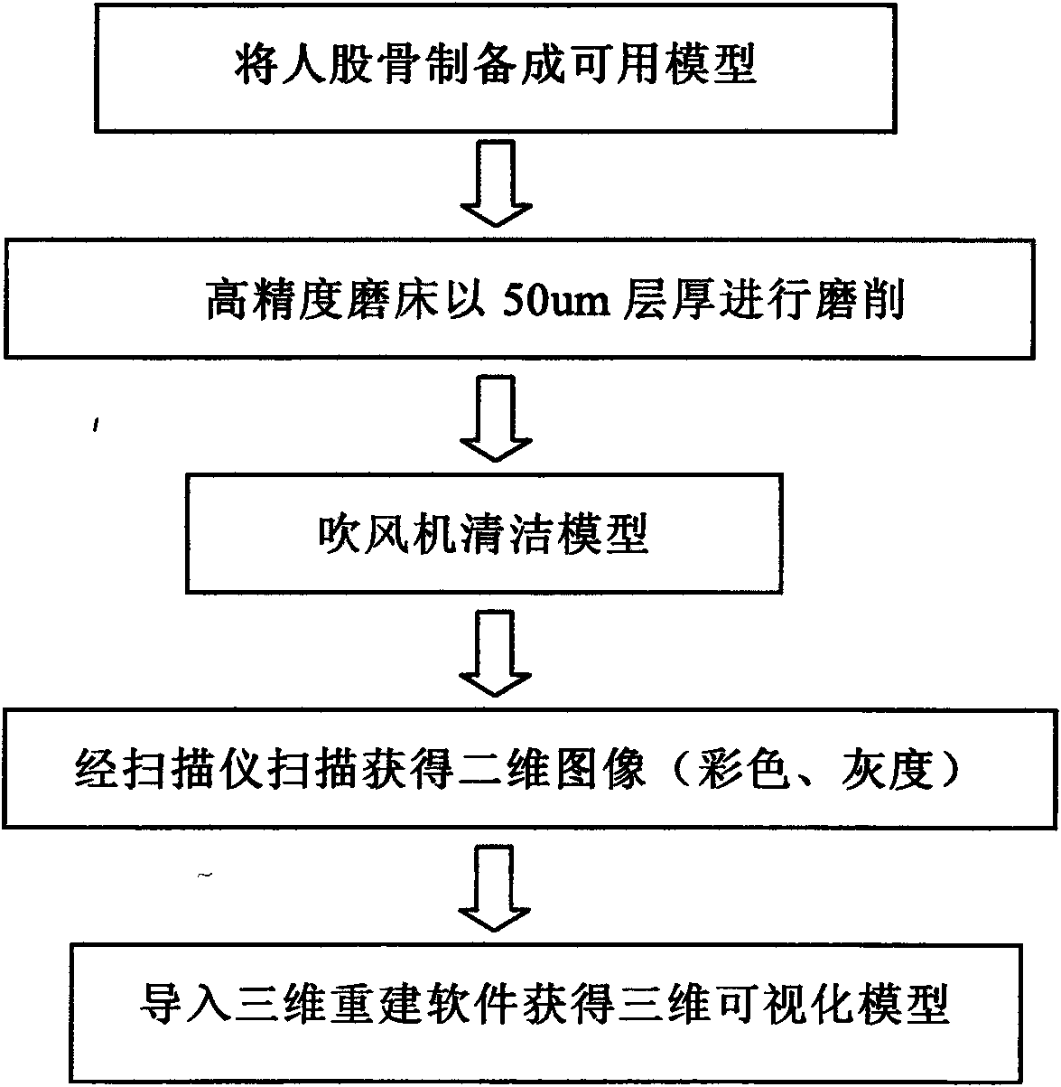
Method for making three-dimensional visualization model of internal structure of bone
A technology of internal structure and production method, which is applied in the field of digital three-dimensional visualization model production of bone internal structure, and can solve problems such as limited application
- Summary
- Abstract
- Description
- Claims
- Application Information
AI Technical Summary
Problems solved by technology
Method used
Image

Examples
Embodiment 1
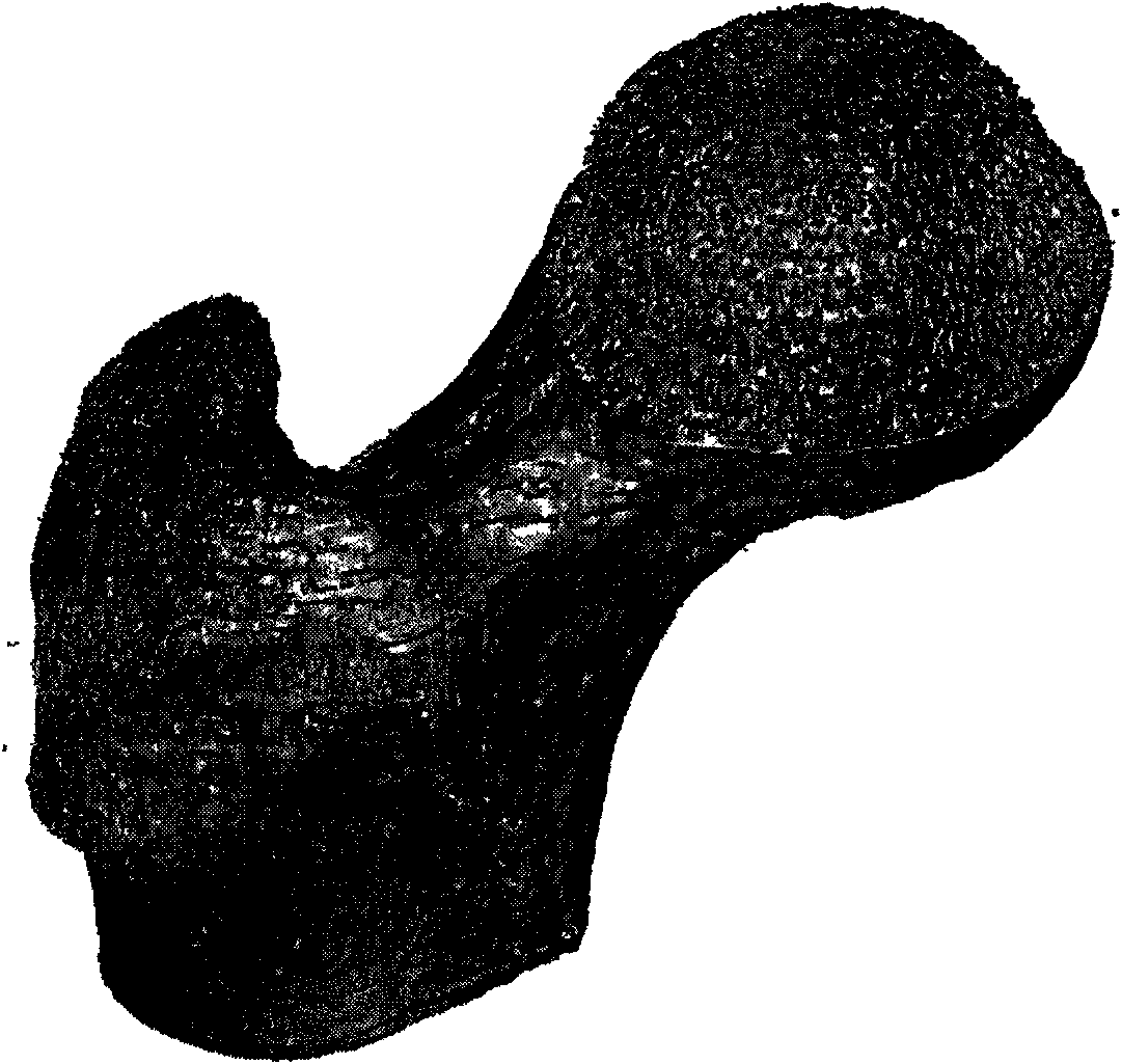
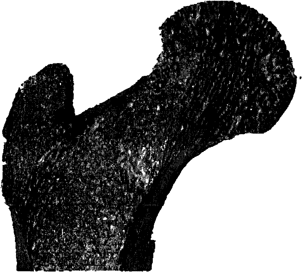
[0019] 1. The present invention selects the side femur of a female corpse who died of cardiovascular disease to prepare a dry bone, and vertically cuts a 150 mm long specimen at the proximal end of the femur, and measures its appearance parameters.
[0020] 2. Take a rectangular polymethyl methacrylate box with a side length of 120 mm and a height of 250 mm as the dry bone specimen container, and the lower part of the dry bone specimen container is fixed on the cast iron base by bonding and Kirschner wires. That is, use chloroform to make an adhesive, firmly bond the bottom of the box body on the square cast iron block with the same cross section, and simultaneously punch holes between the bottom of the box body and the cast iron block (such as 1-3 pieces) of Kirschner wires with a diameter of 2mm to strengthen the fixation. Kirschner wires used for internal fixation in orthopedics generally have a diameter of 0.5-2 mm and a length of about 20 cm. The present invention uses a...
PUM
 Abstract
Description
Claims
Application Information