Kit for testing early lung cancer specific autoantibody enzyme linked immunity and preparation method thereof
An enzyme-linked immunosorbent assay and autoantibody technology, applied in the field of medical detection reagents, can solve problems such as lack of sensitivity and specificity, patient suffering, and inability to diagnose early lung cancer.
- Summary
- Abstract
- Description
- Claims
- Application Information
AI Technical Summary
Problems solved by technology
Method used
Image

Examples
Embodiment 1
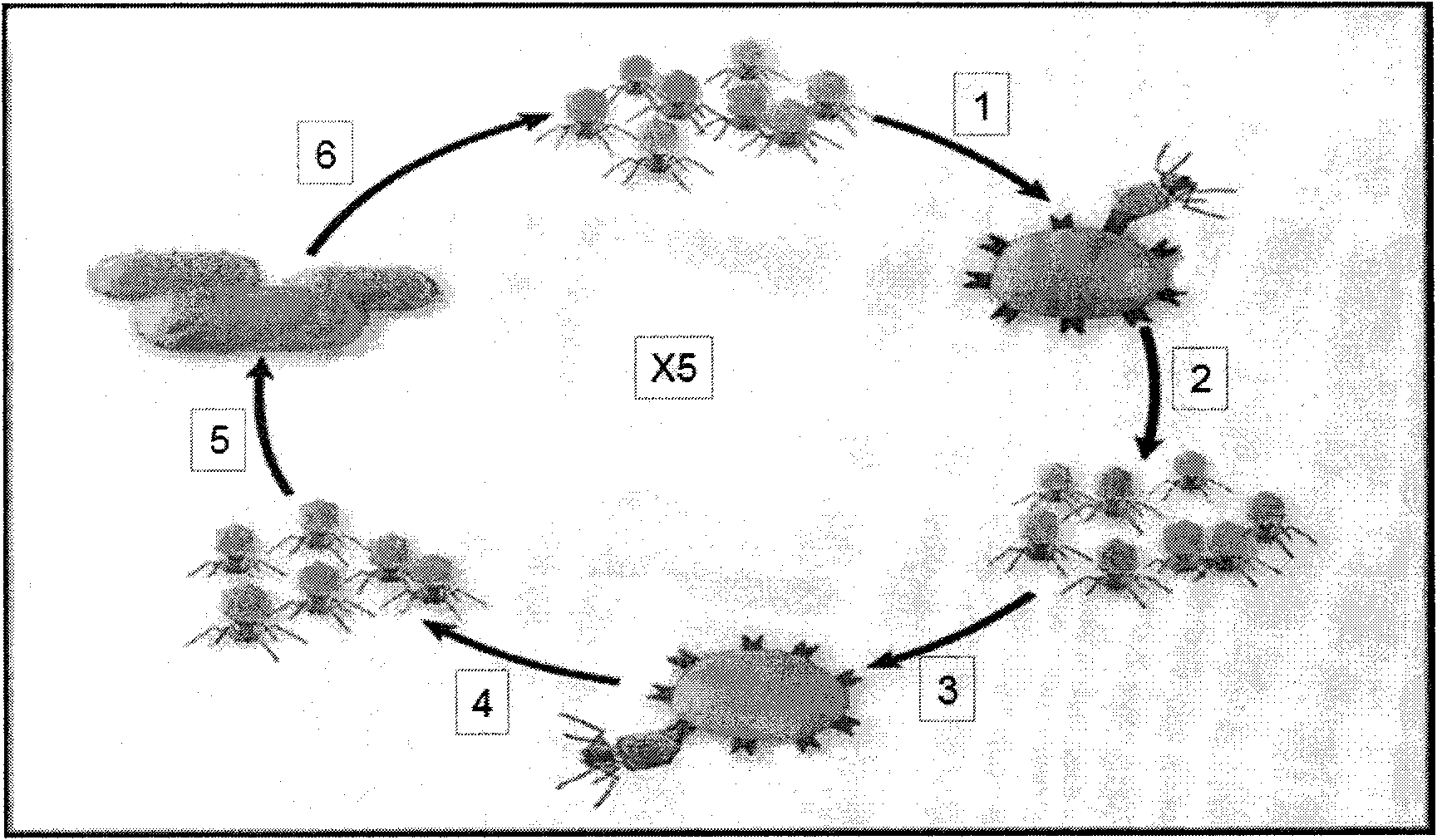
[0049] The T7 tail fiber antibody was diluted 1:1000, each well was coated with 100 μl PBS (PH7.4) on a 96-well ELISA plate, and shaken overnight at 4°C on a shaker. 150 μl washing solution per well, wash four times, about 1 minute each time, and pat dry. 2% BSA / PBS 200ul at room temperature (24°C) to block for 2 hours. ) Add 150 μl washing solution to each well, wash four times, about 1 minute each time, and pat dry. A total of 12 wells were set up, repeated every two wells, and set as auxiliary wells. Add 1% BSA 1:5 diluted lung cancer antigen peptide I (SMMU-EPII) 100 μl to the first and second wells, and add 100 μl to the third and fourth wells 1% BSA 1:5 diluted lung cancer antigen peptide II (SMMU-EPIII) 100μl, lung cancer antigen peptide III (SMMU-EPIIII), lung cancer antigen peptide IV (SMMU-EPIIV), lung cancer antigen peptide V (SMMU-EPIV) and lung cancer Similarly, 100 μl of each antigenic peptide VI (SMMU-EPIVI) was added to the remaining 8 wells sequentially, and...
Embodiment 2
[0051] Coat the T7 tail fiber antibody at a dilution of 1:1000 with 100 μl PBS (PH7.4) per well on a 96-well ELISA plate, shake gently on a shaker at 4°C overnight, add 150 μl washing solution to each well, and wash four times , about 1 minute each time, pat dry. 2% BSA / PBS 200ul at room temperature (24°C) to block for 2 hours. )
[0052] Add 150 μl washing solution to each well, wash four times, about 1 minute each time, and pat dry. A total of 12 wells were set up, and every two wells were repeated, set as auxiliary wells, 1% BSA 1:4 diluted lung cancer antigen peptide I (SMMU-EPII) 100 μl was added to the first and second wells, and 100 μl of lung cancer antigen peptide I (SMMU-EPII) diluted in the third and fourth wells 1% BSA 1:5 diluted lung cancer antigen peptide II (SMMU-EPIII) 100μl, lung cancer antigen peptide III (SMMU-EPIIII), lung cancer antigen peptide IV (SMMU-EPIIV), lung cancer antigen peptide V (SMMU-EPIV) and lung cancer Similarly, 200 μl of each antigeni...
Embodiment 3
[0055] Add 100 μl of 1% BSA 1:500 diluted lung cancer patient plasma to each phage sample, and incubate at room temperature for 1 hour. Wash the plate with washing solution, 150 μl per well, wash four times, about 1 minute each time, and pat dry. Add 100 μl of 1:10000 diluted HRP-coupled goat anti-human IgG to each well and incubate at room temperature for 1 hour. Wash the plate with washing solution, 150 μL per well, wash four times, about 1 minute each time, and pat dry. Add 1 drop each of chromogenic reagents A and B at room temperature to each well, mix well, incubate at 37°C for 20 minutes, and add 25 μl of stop solution. Immediately detect with a microplate reader, take a wavelength of 450nm, first use a blank well to zero, then read the OD value of each well, and record the results.
[0056] Result judgment
[0057] Each ELISA is calculated and judged according to the following formula: after zeroing with a blank well, the sample OD value / the average OD value of the ...
PUM
 Abstract
Description
Claims
Application Information
 - R&D
- Intellectual Property
- Life Sciences
- Materials
- Tech Scout
- Unparalleled Data Quality
- Higher Quality Content
- 60% Fewer Hallucinations
Browse by: Latest US Patents, China's latest patents, Technical Efficacy Thesaurus, Application Domain, Technology Topic, Popular Technical Reports.
© 2025 PatSnap. All rights reserved.Legal|Privacy policy|Modern Slavery Act Transparency Statement|Sitemap|About US| Contact US: help@patsnap.com