Confocal-photoacoustic dual-mode microscopic imaging method and device thereof

A confocal microscopic imaging and microscopic imaging technology, which is used in measurement devices, material analysis by optical means, instruments, etc., can solve the problems of inability to observe cell structure images and low resolution.

Inactive Publication Date: 2010-07-21
SOUTH CHINA NORMAL UNIVERSITY
View PDF5 Cites 18 Cited by
  • Summary
  • Abstract
  • Description
  • Claims
  • Application Information

AI Technical Summary

Problems solved by technology

However, due to the relatively low resolution of current photoacoustic imaging technology, which can onl

Method used


Image

  • Confocal-photoacoustic dual-mode microscopic imaging method and device thereof
  • Confocal-photoacoustic dual-mode microscopic imaging method and device thereof
  • Confocal-photoacoustic dual-mode microscopic imaging method and device thereof

Examples


Embodiment Construction

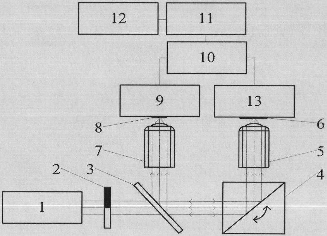
[0021] The present invention will be further described in detail below in conjunction with the embodiments and accompanying drawings, but the embodiments of the present invention are not limited thereto.

[0022] Working process of the present invention is as follows:

[0023] Such as figure 1 As shown, the laser light emitted by the laser 1 is modulated by the chopper 2, and then enters the scanning galvanometer 4 through the half mirror 3 for two-dimensional scanning, and then focuses on the surface of the sample 6 by the microscope objective lens 5. The sample 6 The generated scattered light and fluorescence are collected by the objective lens 5, and after being reflected by the half mirror 3, are focused by the focusing mirror 7 to the pinhole 8, and the scattered light and fluorescence passing through the pinhole 8 are detected by the photomultiplier tube 9, and the photomultiplier After the output signal of the tube 9 is amplified and processed by the lock-in amplifier ...


PUM


Abstract

The invention relates to a confocal-photoacoustic dual-mode microscopic imaging method which comprises the following steps of: (1) carrying out confocal microscopic imaging to an observed object and carrying out photoacoustic scanning imaging to the observed object; and (2) acquiring the functional image of biological tissue by comparing dual-mode images. The invention also relates to a confocal-photoacoustic dual-mode microscopic imaging device which comprises a confocal microscopic imaging system and a photoacoustic scanning imaging system, wherein the confocal microscopic imaging system mainly comprises a laser, a laser scanning galvanometer, a pinhole and a photomultiplier, and the photoacoustic scanning imaging system is formed by sequentially and electrically connecting a photoacoustic sensor, a chopper, a phase-locked amplifier, a data acquisition card and a computer. The invention can carry out confocal-photoacoustic dual-mode microscopic imaging to cells for acquiring the structure information, the content space distribution information and the function information of the cells and the dynamic distribution information of the cell substance contents.

Description

technical field [0001] The invention relates to new technologies of confocal microscopic imaging and photoacoustic microscopic imaging in the field of microscopic imaging, in particular to a confocal-photoacoustic dual-mode microscopic imaging method and a device thereof. Background technique [0002] Confocal microscopy imaging technology has high spatial resolution and unique tomographic imaging capabilities, so it has a wide range of applications in the fields of biomedical research, 3D high-density storage, and 3D microfabrication, and has made many breakthroughs, especially In the field of biomedical research, dynamic high-resolution three-dimensional imaging of tiny objects such as living cells, cell tissues, and even biological macromolecules can be realized, which has become a very effective research method in the field of biomedical research. However, confocal microscopy imaging technology uses scattered light or fluorescence of objects for imaging, and it only obta...

Claims


Application Information

Patent Timeline
IPC IPC(8): G01N21/01G01N29/36
Inventor 唐志列吴泳波廖燕菲
Owner SOUTH CHINA NORMAL UNIVERSITY
Why Eureka
Try Eureka