Application of pumpkin protein in the preparation of drugs for the treatment of lung cancer

A pumpkin and protein technology, applied in the field of anti-tumor drugs, can solve problems such as damage to normal tissues and cells

Inactive Publication Date: 2012-02-15
FUJIAN INST OF RES ON THE STRUCTURE OF MATTER CHINESE ACAD OF SCI +1
View PDF2 Cites 0 Cited by
  • Summary
  • Abstract
  • Description
  • Claims
  • Application Information

AI Technical Summary

Problems solved by technology

[0003] Malignant tumors are diseases that seriously threaten human life and health. Chemotherapy drugs commonly used in clinical practice can greatly damage normal tissues and cells while killing tumor cells, which limits its application.

Method used


Image

  • Application of pumpkin protein in the preparation of drugs for the treatment of lung cancer
  • Application of pumpkin protein in the preparation of drugs for the treatment of lung cancer
  • Application of pumpkin protein in the preparation of drugs for the treatment of lung cancer

Examples


Embodiment 1

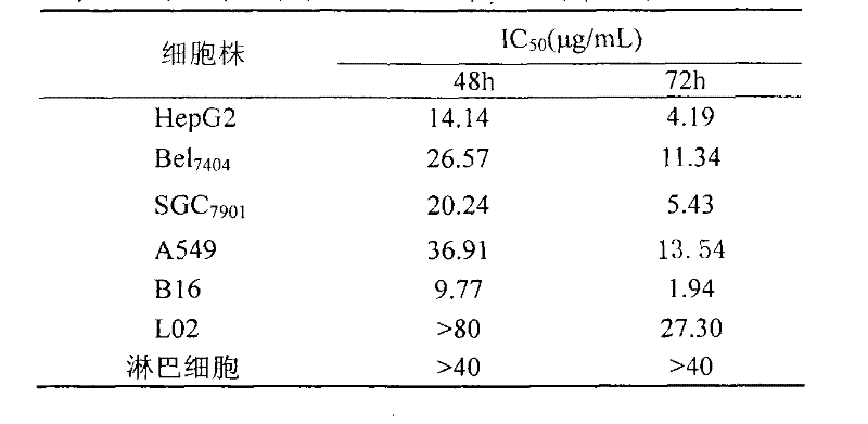
[0011] Example 1: Inhibitory effect of pumpkin protein on the proliferation of tumor cells cultured in vitro and its influence on human normal cells

[0012] Tumor cells in the logarithmic growth phase (gastric cancer cell SGC 7901 , HepG 2 、Bel 7404 , non-small cell lung cancer cell A 549 , breast cancer cells MCF-7 and melanoma cells B16) and normal human liver cells L02 were inoculated on a 96-well culture plate, and the inoculum volume per well was 3000-4000 cells / 100 μL and 6000 cells / 100 μL respectively; healthy blood donations were collected aseptically 15 mL of venous blood from patients, anticoagulated with heparin, separated mononuclear cells by Ficoll density gradient centrifugation, prepared single cell suspension with RPMI1640 containing 10% fetal bovine serum, and adjusted the cell concentration to 2×10 5 cells / mL, inoculated in 96-well culture plate, 100 μL per well. After 24 hours of cell culture on the above-mentioned culture plates, 100 μL of pumpkin prot...

Embodiment 2

[0015] Example 2: Pumpkin protein to SGC 7901 , HepG 2 、Bel 7404 、A 549 , MCF-7 and B16 cells inducing apoptosis

[0016] (1) Effect of pumpkin protein on the morphology of the above tumor cells

[0017] ①Fluorescence microscope observation: the above tumor cells were inoculated on multiple coverslips, treated with different concentrations of pumpkin protein for 24, 48 and 72 hours after 12 hours, and stained with acridine orange (AO) / ethidium bromide (EB) mixed fluorescent dyes Afterwards, place it upside down on a glass slide, and observe the morphological changes of the cells under a fluorescence microscope. Results: The above cells can be observed in the nucleus or cytoplasm of apoptotic cells in the early and middle stages, with densely stained granular yellow-green fluorescence, nuclear chromatin condensation, nuclear rupture, and the release of apoptotic bodies. Typical apoptotic cell morphology There are time- and dose-dependent changes in apoptosis.

[0018] ② E...

Embodiment 3

[0022] Example 3: Induction and differentiation of pumpkin protein on tumor cells (using melanoma B16 cells as a model)

[0023] (1) Observation of cell morphology: After the B16 cells in the control group and the experimental group were cultured for 72 hours, the cell morphology was observed and photographed with a phase contrast microscope and a transmission electron microscope, respectively. The result is as follows:

[0024] ①Observation by optical microscope: B16 cells in the control group grew irregularly in multiple layers, with overlapping round cells; after the treatment of pumpkin protein, the cells grew in a certain polarity, arranged in parallel without overlapping; after 3 days, the concentration of pumpkin protein was 2.5 For those above μg / mL, the polarity of the cells is obvious, most of the cells become larger, have a dendritic structure, the cells are connected into a network, grow slowly, and the number of cells decreases.

[0025] ② Electron microscope obser...


PUM

PropertyMeasurementUnit
molecular weightaaaaaaaaaa

Abstract

The application of pumpkin protein in pharmacy can isolate a ribosome inactivating protein-pumpkin protein from pumpkin pulp, and the protein shows good antitumor activity and can be used to prepare antitumor drugs. Pumpkin protein can significantly inhibit gastric cancer cells SGC7901 cultured in vitro, liver cancer cells HepG2, Bel7404, non-small cell lung cancer cells A549, breast cancer cells MCF-7 and melanoma cells B16, while inhibiting human normal liver cells and peripheral blood lymphocytes. The effect is small, showing obvious selectivity; pumpkin protein can also significantly inhibit cervical cancer U14, melanoma B16 and lung cancer Lewis and other animal transplanted tumors in mice. In addition, the present invention also confirms that the apoptosis-inducing and differentiation-inducing effect of pumpkin protein on tumor cells is an important mechanism of its anti-tumor activity.

Description

[0001] This application is a divisional application of the invention patent: application of pumpkin protein in pharmaceuticals (application date: November 3, 2005; application number: 200510119741.2). technical field [0002] The invention relates to the field of antitumor drugs, in particular to the application of pumpkin protein in the preparation of antitumor drugs. Background technique [0003] Malignant tumors are diseases that seriously threaten human life and health. Chemotherapy drugs commonly used in clinical practice can greatly damage normal tissues and cells while killing tumor cells, which limits its application. It has become the direction and urgent task of current anticancer drug research to find highly efficient and low toxic drugs for treating tumors. Finding anti-tumor drugs from natural substances has been considered an effective way, and the research on the anti-tumor effects of natural active substances, especially in the aspects of inhibiting prolifera...

Claims


Application Information

Patent Timeline
Patent Type & Authority Patents(China)
IPC IPC(8): A61K38/16A61P35/00A61P11/00
Inventor 陈明晃谢捷明
Owner FUJIAN INST OF RES ON THE STRUCTURE OF MATTER CHINESE ACAD OF SCI
Why Eureka
Try Eureka