Human embryonic trigeminus based three-dimensional reconstruction method by using histotomy staining

A trigeminal nerve and three-dimensional reconstruction technology, which is applied in 3D modeling, test sample preparation, image data processing, etc., can solve the problem of no human embryo trigeminal nerve root, etc., and achieve the effect of specific, feasible, simple and practical methods

Inactive Publication Date: 2010-12-15
XINJIANG MEDICAL UNIV
View PDF4 Cites 26 Cited by
  • Summary
  • Abstract
  • Description
  • Claims
  • Application Information

AI Technical Summary

Problems solved by technology

[0005] There is no research report on the course of human embryonic trigeminal nerve root and motor root

Method used


Image

  • Human embryonic trigeminus based three-dimensional reconstruction method by using histotomy staining
  • Human embryonic trigeminus based three-dimensional reconstruction method by using histotomy staining
  • Human embryonic trigeminus based three-dimensional reconstruction method by using histotomy staining

Examples


Embodiment 1

[0034] Example 1: Three-dimensional reconstruction method based on human embryonic trigeminal nerve using tissue section staining

[0035] 1. Specimen fixation

[0036] Within 1-4 hours after obtaining the specimen, clamp the fetal umbilical cord with forceps, open the anterior chest wall, expose the pericardium, cut the pericardium, fully expose the heart, insert a No. 7 infusion needle into the root of the ascending aorta, and cut the right atrium with scissors Make a small incision, use a 50ml syringe to continuously inject 500ml of 0.9% normal saline to wash the heart, and then inject 1500ml of 10% formalin solution, let the fixative perfusion fix along the systemic circulation, observe the fixation effect of the specimen, see The fixative fluid flows out from the mouth, nose, etc. of the specimen, the abdomen of the specimen swells, and the limbs become hard, indicating that the fixation effect of the specimen is good. Continue to soak the perfused specimen in freshly pre...

Embodiment 2

[0063] Embodiment 2: Specimen fixation

[0064] Within 1-4 hours after obtaining the specimen, clamp the fetal umbilical cord with forceps, open the anterior chest wall, expose the pericardium, cut the pericardium, fully expose the heart, insert a No. 7 infusion needle into the root of the ascending aorta, and cut the right atrium with scissors Make a small incision, use a 50ml syringe to continuously inject 500ml of 0.9% normal saline to wash the heart, and then inject 1500ml of 10% formalin solution, let the fixative perfusion fix along the systemic circulation, observe the fixation effect of the specimen, see The fixative fluid flows out from the mouth, nose, etc. of the specimen, the abdomen of the specimen swells, and the limbs become hard, indicating that the fixation effect of the specimen is good. Continue to soak the perfused specimen in freshly prepared 10% formalin solution for post-fixation, and place it in a dry place. Keep away from light.

Embodiment 3

[0065] Embodiment three: light microscope, transmission electron microscope observation

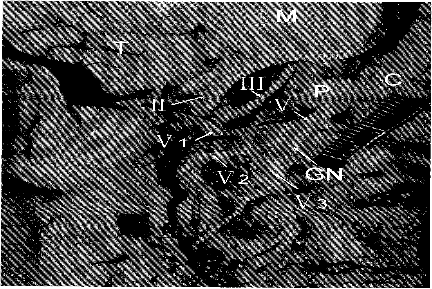
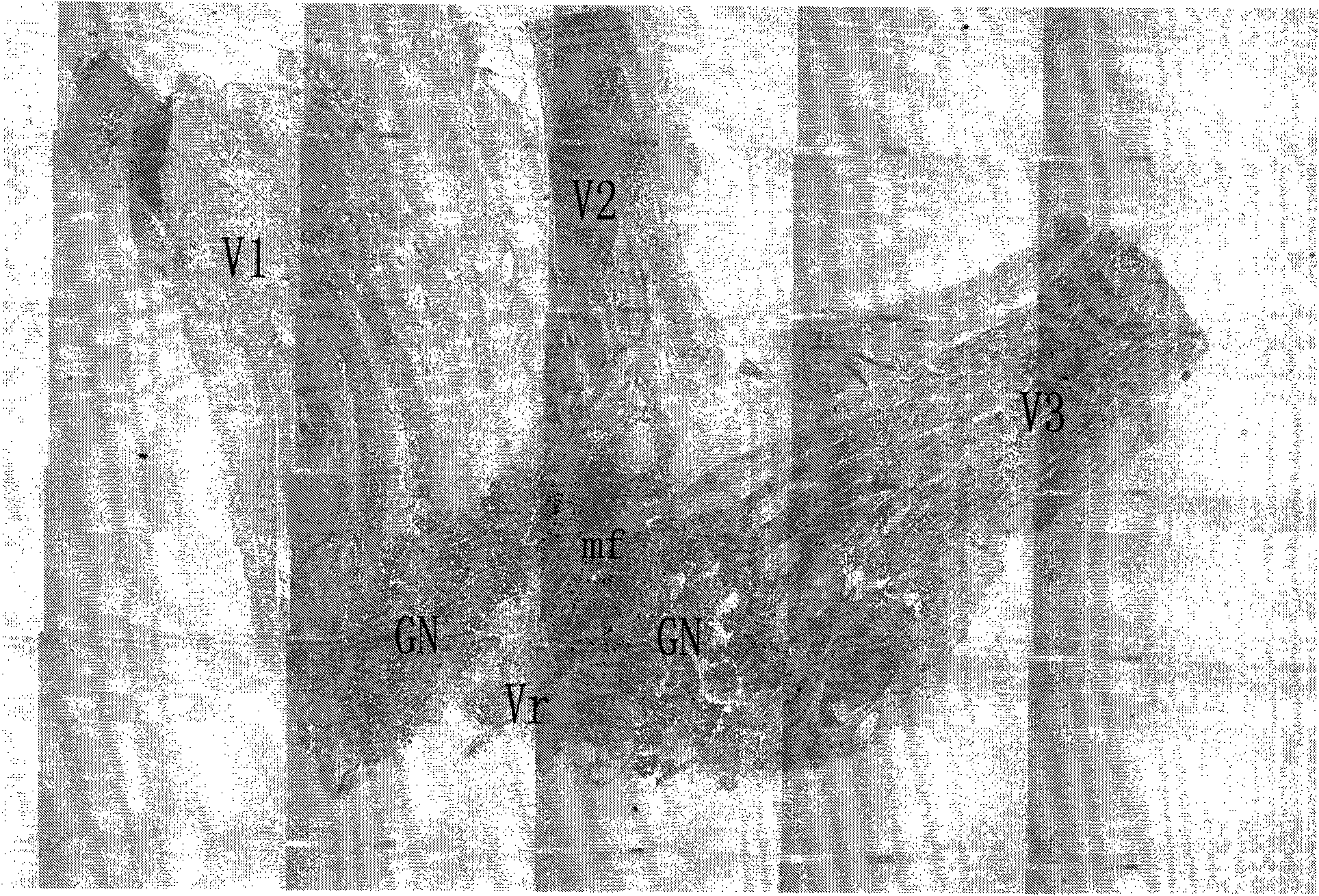
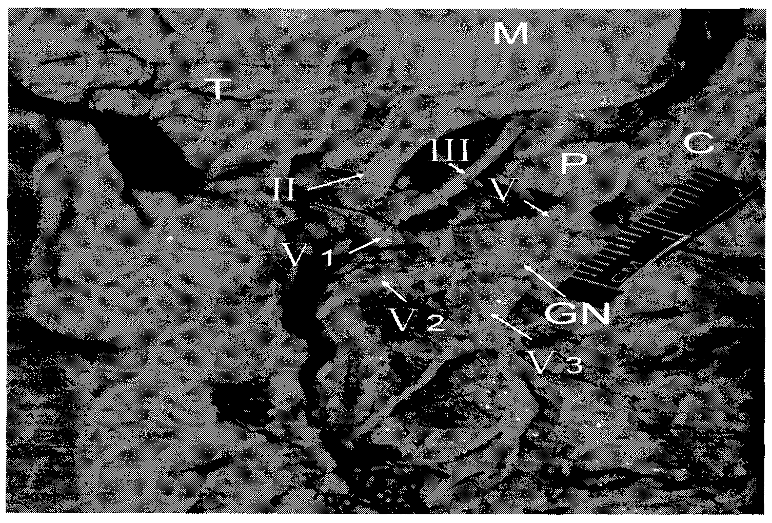
[0066] (1) Material collection: The right trigeminal nerve root, trigeminal ganglion and three branches of the right trigeminal nerve root, trigeminal ganglion and three branches were completely removed from the specimens in the above examples. After trimming the tissue, the motor root of the trigeminal nerve can be distinguished with the naked eye. Motor nerve roots of nerve segments, see appendix figure 1 ,2 .

[0067] (2) Pre-fixation: the taken tissues were marked and placed in containers filled with 2.5% glutaraldehyde solution, and the fixation time was 72 hours.

[0068] (3) Rinsing: Take out the fixed and marked tissue, transfer to 1 / 15M phosphate buffer and rinse in pH 7.4 for 3 times, each time for 15 minutes.

[0069] (4) Post-fixation: Post-fix the rinsed tissues for 1 hour in 1% HCl fixative.

[0070] (5) Rinsing: the operation steps are the same as (3).

[0071] (6) Dehy...


PUM

PropertyMeasurementUnit
thicknessaaaaaaaaaa

Abstract

The invention discloses a human embryonic trigeminus based three-dimensional reconstruction method by using histotomy staining. In the method, trigeminal ganglions, roots and branches of a fresh sample of a human embryo, especially an over 20-week induced-labor infant embryo not died with diseases are selected and subjected to HE conventional staining and observed under a light microscope and a transmission electron microscope, and finally the three-dimensional reconstruction is realized by using a computer TRI for win 95 / NT, Version:3.01. By researching the form of the human embryonic trigeminus and advancing of motor roots, the invention shows the three-dimensional structure of the human embryonic trigeminus for the first time in human history, which not only enriches the research content of morphology of human embryonic trigeminus, but also provides a morphological basis for clinical prosopalgia pathophysiology and treatment. And thus, the human embryonic trigeminus based three-dimensional reconstruction method by using histotomy staining has a wide application scope.

Description

technical field [0001] The invention belongs to the technical field of neuroanatomy. Specifically, the present invention relates to a technical field of three-dimensional reconstruction based on human embryonic trigeminal nerve using tissue section staining. Background technique [0002] The trigeminal nerve is a thick mixed cranial nerve that contains both general somatosensory and specific visceral motor fibers. Special visceral motor fibers originate from the motor nucleus of the trigeminal nerve in the middle part of the pons, and its axons form the motor root of the trigeminal nerve. The mandibular nerve exits the cranium through the foramen ovale and distributes to the muscles of mastication along with the branches of the mandibular nerve. The cell bodies of general somatosensory fibers are concentrated in the trigeminal ganglion, in which the central process is concentrated to form a thick trigeminal sensory root, which enters the brain from the junction of the pont...

Claims


Application Information

Patent Timeline
Patent Type & Authority Applications(China)
IPC IPC(8): G01N1/30G06T17/00
Inventor 古丽尼沙·克力木栾新平吴励郭莲萍吐尔逊江·达地汗迪力夏提·艾尼廖礼彬德力夏提·依米提毛新民王蕾王水泉贠江利陈荣胡汉华王士平
Owner XINJIANG MEDICAL UNIV
Why Eureka
Try Eureka