Human embryonic trigeminus based three-dimensional reconstruction method by using slide stainer
A trigeminal nerve and three-dimensional reconstruction technology, which is applied in 3D modeling, test sample preparation, image data processing, etc., can solve the problem of no human embryo trigeminal nerve root, etc., and achieve the effect of specific, feasible, simple and practical methods
- Summary
- Abstract
- Description
- Claims
- Application Information
AI Technical Summary
Problems solved by technology
Method used
Image
Examples
Embodiment 1
[0034] Example 1: Three-dimensional reconstruction method based on human embryonic trigeminal nerve using tissue section staining
[0035] 1. Specimen fixation
[0036] Within 1-4 hours after obtaining the specimen, clamp the fetal umbilical cord with forceps, open the anterior chest wall, expose the pericardium, cut the pericardium, fully expose the heart, insert a No. 7 infusion needle into the root of the ascending aorta, and cut the right atrium with scissors Make a small incision, use a 50ml syringe to continuously inject 500ml of 0.9% normal saline to wash the heart, and then inject 1500ml of 10% formalin solution, let the fixative perfusion fix along the systemic circulation, observe the fixation effect of the specimen, see The fixative fluid flows out from the mouth, nose, etc. of the specimen, the abdomen of the specimen swells, and the limbs become hard, indicating that the fixation effect of the specimen is good. Continue to soak the perfused specimen in freshly pre...
Embodiment 2
[0063] Embodiment 2: Specimen fixation
[0064] Within 1-4 hours after obtaining the specimen, clamp the fetal umbilical cord with forceps, open the anterior chest wall, expose the pericardium, cut the pericardium, fully expose the heart, insert a No. 7 infusion needle into the root of the ascending aorta, and cut the right atrium with scissors Make a small incision, use a 50ml syringe to continuously inject 500ml of 0.9% normal saline to wash the heart, and then inject 1500ml of 10% formalin solution, let the fixative perfusion fix along the systemic circulation, observe the fixation effect of the specimen, see The fixative fluid flows out from the mouth, nose, etc. of the specimen, the abdomen of the specimen swells, and the limbs become hard, indicating that the fixation effect of the specimen is good. Continue to soak the perfused specimen in freshly prepared 10% formalin solution for post-fixation, and place it in a dry place. Keep away from light.
Embodiment 3
[0065] Embodiment three: light microscope, transmission electron microscope observation
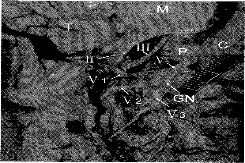
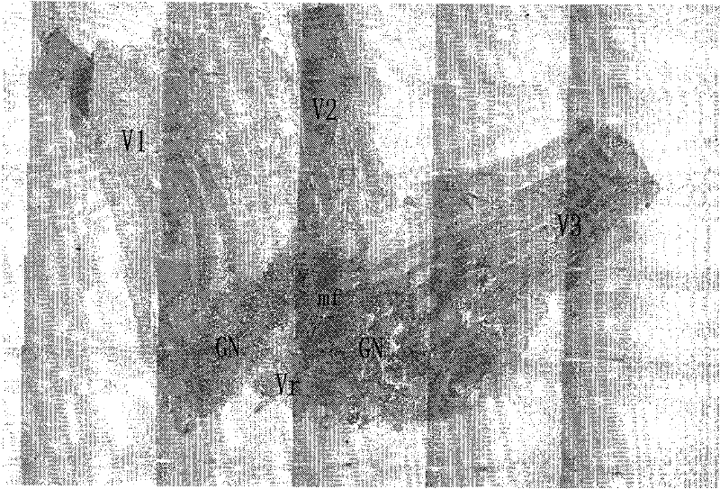
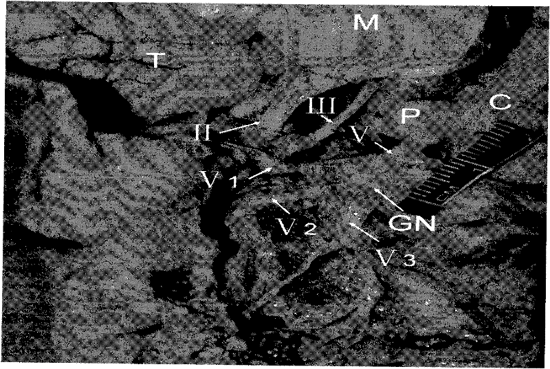
[0066] (1) Material collection: The right trigeminal nerve root, trigeminal ganglion and three branches of the right trigeminal nerve root, trigeminal ganglion and three branches were completely removed from the specimens in the above examples. After trimming the tissue, the motor root of the trigeminal nerve can be distinguished with the naked eye. Motor nerve roots of nerve segments, see appendix figure 1 ,2 .
[0067] (2) Pre-fixation: the taken tissues were marked and placed in containers filled with 2.5% glutaraldehyde solution, and the fixation time was 72 hours.
[0068] (3) Rinsing: Take out the fixed and marked tissue, transfer to 1 / 15M phosphate buffer and rinse in pH 7.4 for 3 times, each time for 15 minutes.
[0069] (4) Post-fixation: Post-fix the rinsed tissues for 1 hour in 1% HCl fixative.
[0070] (5) Rinsing: the operation steps are the same as (3).
[0071] (6) Dehy...
PUM
| Property | Measurement | Unit |
|---|---|---|
| thickness | aaaaa | aaaaa |
Abstract
Description
Claims
Application Information
 - R&D
- Intellectual Property
- Life Sciences
- Materials
- Tech Scout
- Unparalleled Data Quality
- Higher Quality Content
- 60% Fewer Hallucinations
Browse by: Latest US Patents, China's latest patents, Technical Efficacy Thesaurus, Application Domain, Technology Topic, Popular Technical Reports.
© 2025 PatSnap. All rights reserved.Legal|Privacy policy|Modern Slavery Act Transparency Statement|Sitemap|About US| Contact US: help@patsnap.com


