In-vitro micro organ and related use thereof

A technology of micro-organs and organs, applied in the direction of using vectors to introduce foreign genetic material, cells modified by introducing foreign genetic material, recombinant DNA technology, etc., which can solve problems such as low shear stress

Inactive Publication Date: 2011-04-13
YISSUM RES DEV CO OF THE HEBREWUNIVERSITY OF JERUSALEM LTD
View PDF20 Cites 5 Cited by
  • Summary
  • Abstract
  • Description
  • Claims
  • Application Information

AI Technical Summary

Problems solved by technology

In summary, conventional culture methods cannot simultaneously achieve sufficiently low shear stress, sufficient three-dimensional spatial freedom, and critical cell interactions (with each other or the substrate) for a sufficiently long period to allow excellent modeling of tissue architecture in vivo

Method used


Image

  • In-vitro micro organ and related use thereof
  • In-vitro micro organ and related use thereof
  • In-vitro micro organ and related use thereof

Examples


Embodiment

[0212] The invention, now being progressively described, will be more readily understood by reference to the following examples, which are included solely for the purpose of illustrating certain aspects and embodiments of the invention, and are not intended to limit the invention.

[0213] Microorgan cultures from animals including human adult skin, mouse, guinea pig and rat skin have been isolated and grown in culture for up to 21 days. However, it is also within the scope of the invention that the culture be maintained for a period of time greater than 21 days.

[0214] In addition, it is within the scope of the invention to form micro-organ cultures from various animals. The range of animals is exemplary only, but not limited to the samples provided below.

[0215]As described in the appended examples, micro-organ cultures were prepared from skin and from organs including mammalian pancreas, liver, kidney, duodenum, esophagus and bladder. Similarly, micro-organ cultures f...

Embodiment I

[0218] Preparation of epidermal micro-organ cultures

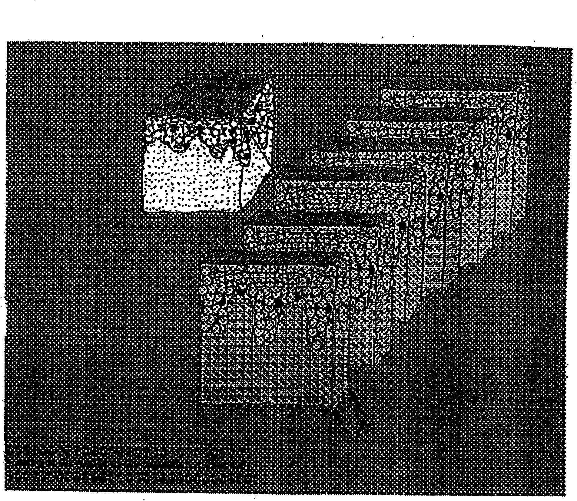
[0219] Fresh skin was obtained after surgery, and the underlying fatty tissue was removed and cut into 0.4×5 cm flaps, and then cut transversely into 300 μm slices under sterile conditions using a tissue chopper or other suitable cutting tools, so that the final Tissue segments have a size of 4 mm in width and 0.3 mm in thickness (see figure 1 ). These micro-organs were placed in 24-well microplates in 400 μl DMEM without serum, 5% CO 2 , 37° C., with continuous shaking at 12 rpm for one to eight days. Twenty micro-explants were grown per well.

Embodiment 2

[0221] Determination of proliferation in mouse, guinea pig, and human epidermal micro-organ cultures

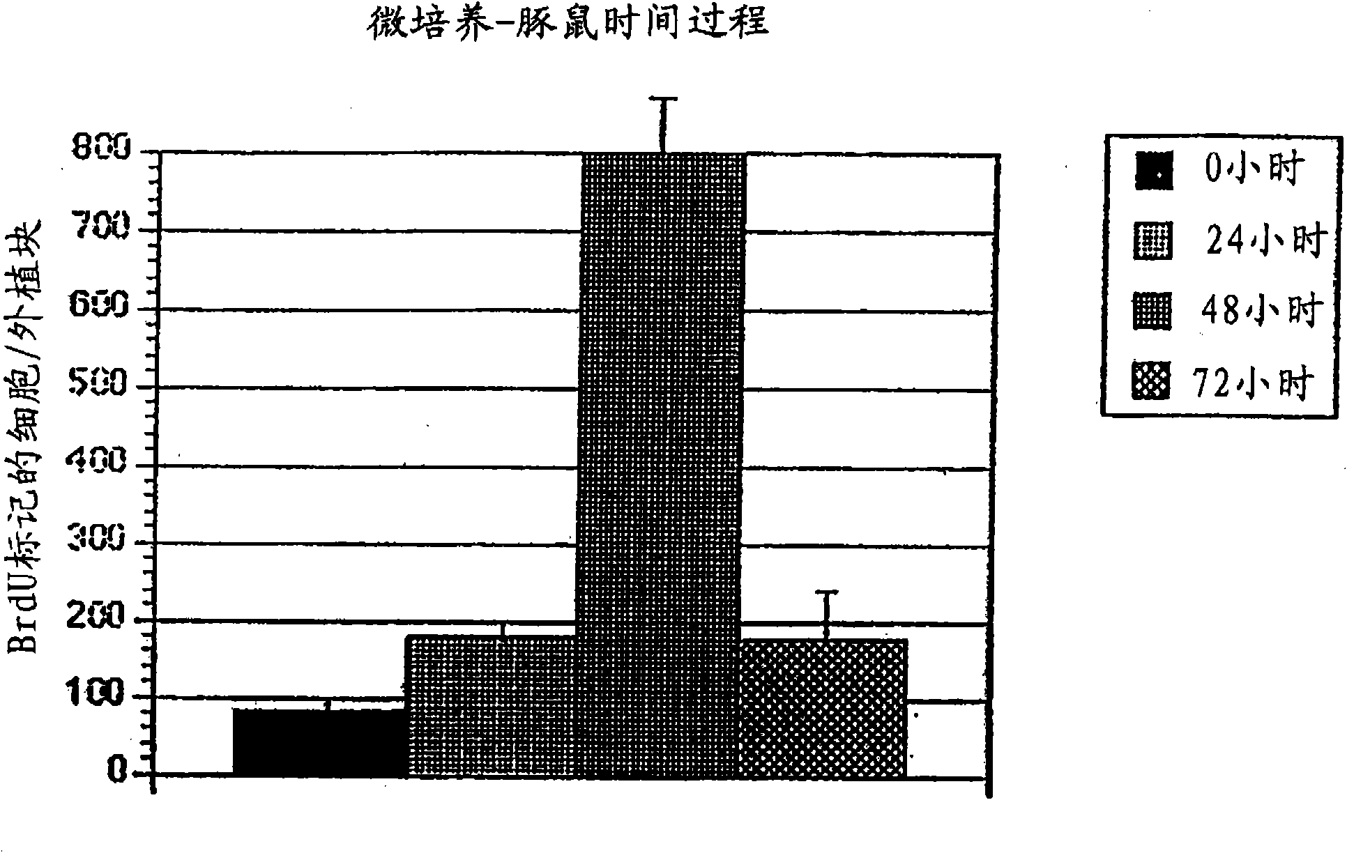
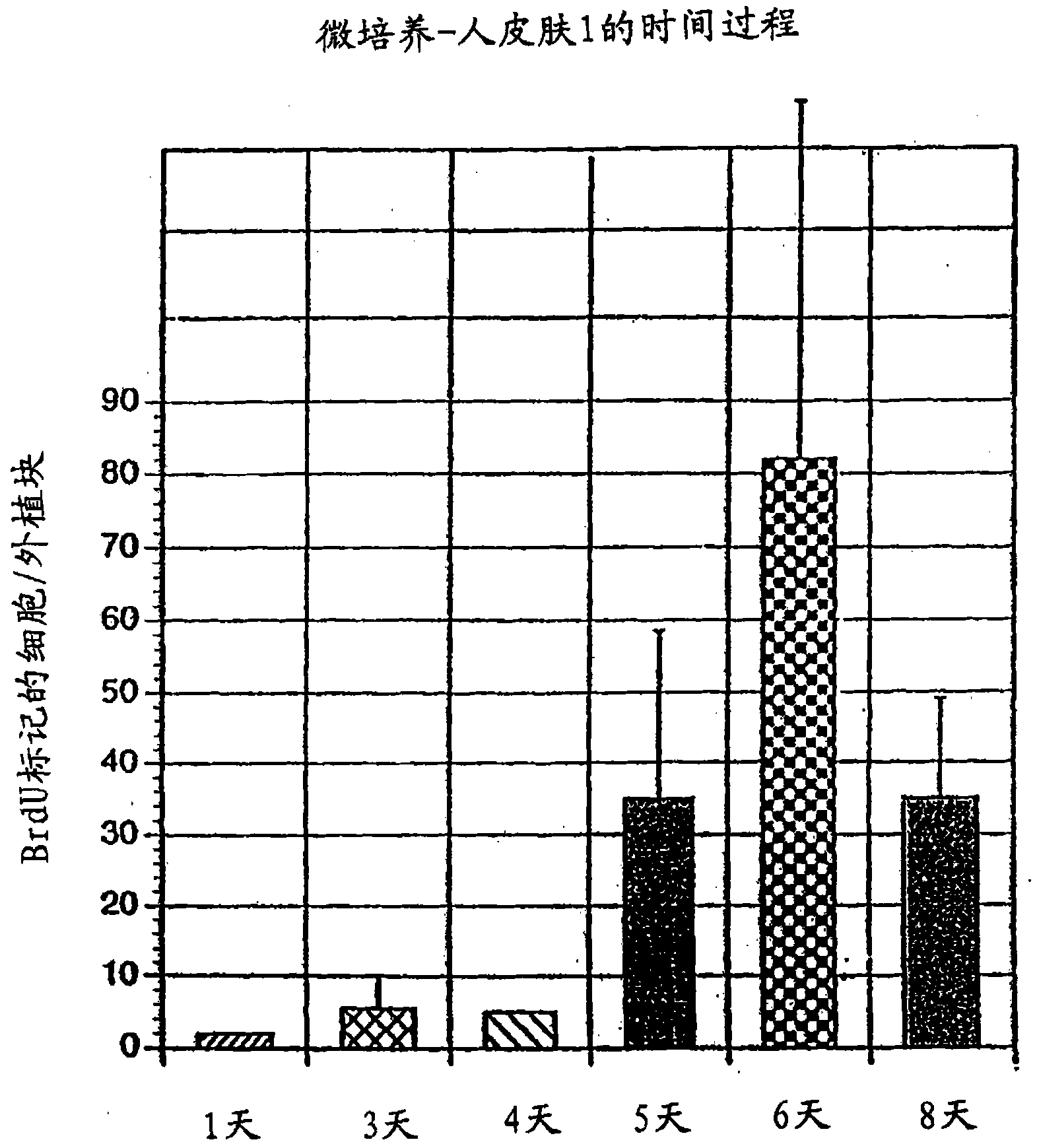
[0222] Micro-organ cultures were prepared according to Example I, and the amount of DNA synthesis was analyzed to determine cell proliferation as follows. Mouse skin and guinea pig skin were grown for two days and human skin for four days, after which BrdU was added to the medium at a final concentration of 100 μΜ for three hours, and cells were subsequently fixed in 4% formaldehyde. After fixation, cultures were stained with goat anti-BrdU antibody followed by anti-goat FICT-labeled IgG. Histological specimens were embedded in 4% formaldehyde in the following fixatives and sectioned at 3 μm and stained with methylene blue.

[0223] It was found that after two to four days of culture in vitro, the fraction of cells synthesizing DNA increased up to 10-fold compared to the value observed in vivo, after which the rate of DNA synthesis gradually increased but was maintained up t...


PUM

PropertyMeasurementUnit
diameteraaaaaaaaaa

Abstract

The invention discloses a micro organ culture for separated cell population with special characteristics. The topic micro organ culture has the prominent characteristics of capacity of maintaining a relatively long period in the culture and an organ micro-architecture for facilitating cell-cell and cell-matrix interaction present in a source organ. The micro organ culture can be used in a method for transferring gene products to a receptor subject to identify hyperplasia and cell differentiation reagents and identify and separate progenitor cells from stem cells. In addition, the micro organ culture can be used in a method for identifying hyperplasia, cell differentiation and virus infectious inhibitors. In other schemes, the micro organ culture can be used for transplant.

Description

[0001] This application is a divisional application of the Chinese patent application with the application number 01823906.4, the application date is October 23, 2001, and the invention title is "in vitro micro-organs and related applications". technical field [0002] The present invention relates to in vitro micro-organs and related uses thereof. Background technique [0003] Eukaryotic cell culture was accomplished in the early 1950s. Since then, a wide range of transformed and primary cells have been cultured using a wide variety of media with well-defined supplements such as growth factors and hormones, and less well-defined supplements such as serum and other body extracts. For example, fibroblasts derived from animal skin can be routinely cultured through many cell passages such as karyotype diploid cells or infinitely such as established cell lines. However, epithelial cells have different morphological and proliferative properties than fibroblasts and are more diff...

Claims


Application Information

Patent Timeline
Patent Type & Authority Applications(China)
IPC IPC(8): C12N5/10C12N15/86A61K35/12
Inventor E·N·米特拉尼
Owner YISSUM RES DEV CO OF THE HEBREWUNIVERSITY OF JERUSALEM LTD
Why Eureka
Try Eureka