Kit for detecting free breast cancer cell marker in blood

A breast cancer cell and kit technology, applied in measurement devices, instruments, scientific instruments, etc., can solve the problems that ordinary patients are difficult to accept, tumor cells are not specific, and detection methods are inaccurate.

Active Publication Date: 2011-06-29
牛刚 +2
View PDF5 Cites 5 Cited by
  • Summary
  • Abstract
  • Description
  • Claims
  • Application Information

AI Technical Summary

Problems solved by technology

Second, the chemical and physical means adopted have no therapeutic specificity for tumor cells
But this detection method is very inaccurate
We know that the increase in the content of tumor markers may be due to the secretion of tumor cells, or other factors such as stress responses may cause the increase in the protein content of the marker, so even if the increase in the content of tumor markers is detected, it cannot be accurately...

Method used


Image

  • Kit for detecting free breast cancer cell marker in blood
  • Kit for detecting free breast cancer cell marker in blood
  • Kit for detecting free breast cancer cell marker in blood

Examples


Embodiment 1

[0039] The preparation of embodiment 1 magnetic beads

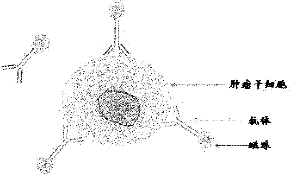
[0040] Preparation of antibody immunomagnetic beads: the antibodies used were anti-BerEP4 monoclonal antibody, anti-cytokeratin monoclonal antibody and anti-HER2 monoclonal antibody. In the present invention, the above three kinds of antibodies are labeled separately and then mixed for use. It is also possible to select one or two antibodies for separate labeling and mixed use.

[0041] Methods: Dynabeads Antibody Coupling Kit produced by Invitrogen was used. The labeling method was carried out in strict accordance with the manufacturer's instructions.

Embodiment 2

[0042] The separation of embodiment 2 breast cancer cells

[0043] 1. Sample collection:

[0044] Collect 5-7.5ml of peripheral blood from breast cancer patients, anticoagulate with EDTA, and use it within 4 hours (or use it within 48 hours after storage at 4 degrees).

[0045] 2. Select magnetic beads:

[0046] 2.1 Handling of magnetic beads:

[0047] Mix the magnetic beads evenly, use a pipette to blow gently, and do not use a vortexer;

[0048] Use PBS buffer solution to wash, and do not touch the microspheres when washing;

[0049] Draw more magnetic beads than the required sample volume and add them to a 1.5ml centrifuge tube;

[0050] Put it on the magnet for 1 minute, discard the supernatant;

[0051] Wash 3 times with 1ml PBS to remove preservatives;

[0052] Remove the supernatant;

[0053] · Aspirate 100ul microspheres containing PBS with the same volume as the original one.

[0054] 2.2 Selection of tumor cells

[0055] Put 5ml of peripheral blood into a 15...

Embodiment 3

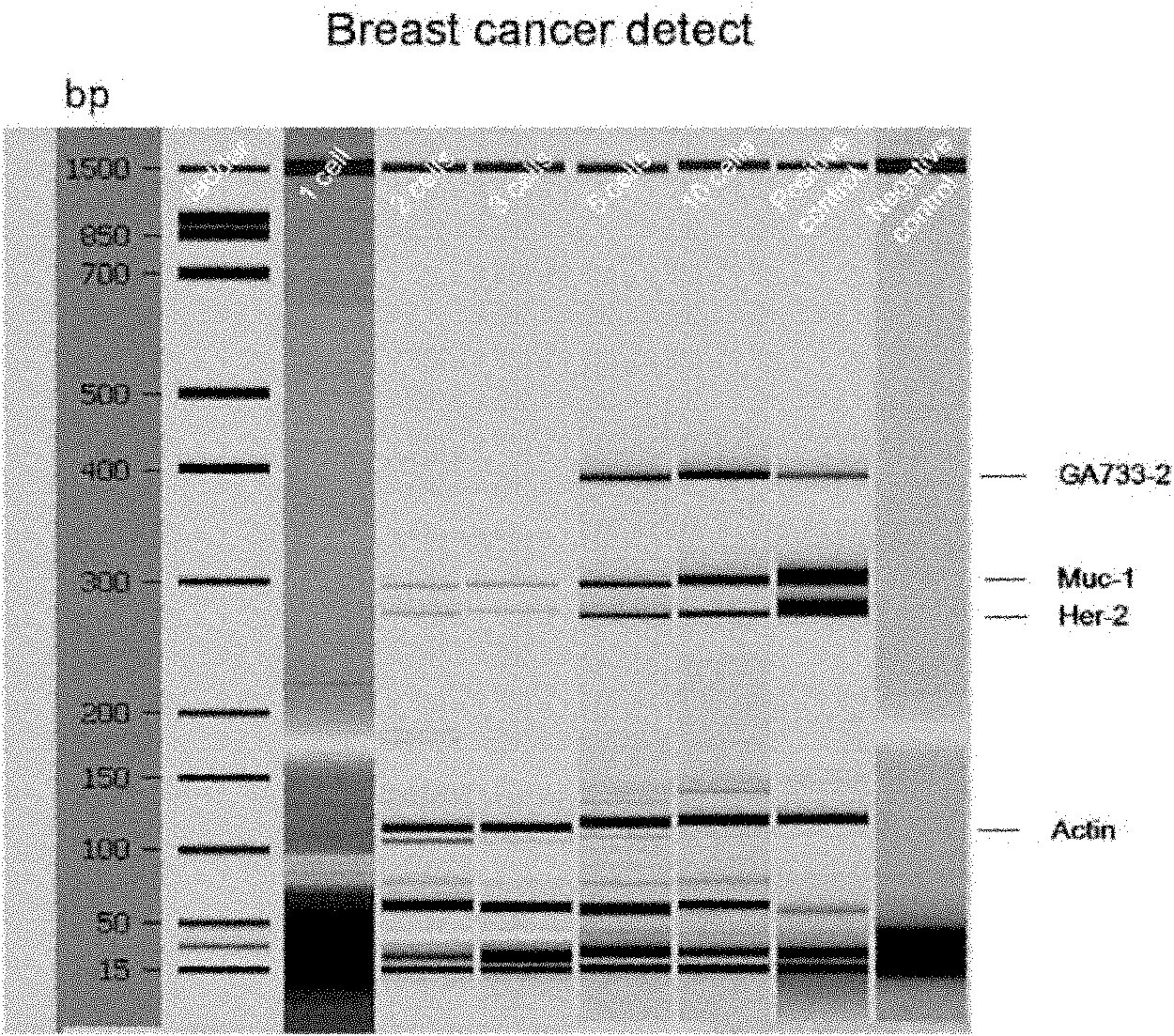
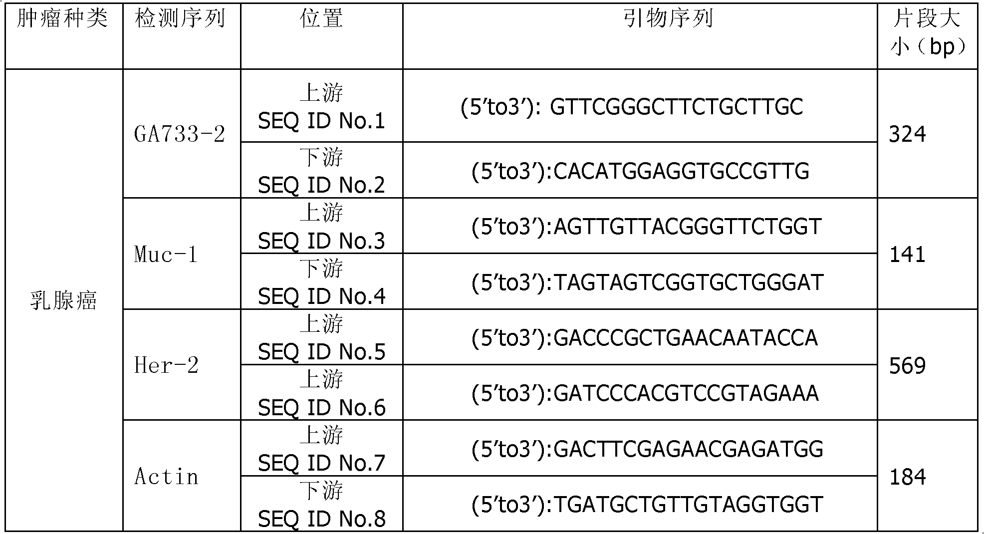
[0074] Example 3 Detection of Breast Cancer Cell Tumor Markers

[0075] 3.1 Kit preparation

[0076] Keep the test tube at room temperature;

[0077] Put the RNase-free water in the kit at room temperature;

[0078] Place the centrifuge tube above the tube containing the supernatant on ice;

[0079] · Prepare magnetic beads.

[0080] 3.2 Preparation of oligonucleotides

[0081] Take an appropriate amount of magnetic beads with oligonucleotide Oligo(dT) (it is recommended not to use a suspension apparatus to mix, but to suspend by hand);

[0082] · Transfer to a 1.5ml tube;

[0083] • Wash twice with lysis / binding buffer.

[0084] 3.3 Binding mRNA to magnetic beads

[0085] Add 20 μl of magnetic beads to each cell lysis sample;

[0086] • Incubate for 10 minutes.

[0087] 3.4 Purification of mRNA

[0088] Wash twice with buffer solution;

[0089] Wash the magnetic beads twice with buffer;

[0090] Wash again with 100 μl Tris-HCl buffer;

[0091] • Suspend the magne...


PUM


Abstract

The invention discloses a kit for detecting a breast cancer marker. The kit comprises the following components: magnetic bead processing solution, a magnetic bead with a breast cancer antibody, 4.0 to 5.0mu l of 10X buffer solution, 4.0 to 5.0mu l of deoxynucleoside triphosphates (dNTPs), 2.0 to 3.0mu l of reverse transcriptase, 0.4 to 0.6mu l of 40u/mu l ribonucleic acid (RNA) enzyme inhibitor, 20.0 to 30.0mu l of 10X hot start polymerase chain reaction (PCR) mixed solution, 10.0 to 15.0mu l of ddH2O and 3.0 to 5.0mu l of breast cancer marker primer. Compared with the prior art, the kit can more accurately detect the breast cancer marker in peripheral blood, and has great guiding significance for selecting breast cancer prognosis schemes and treatment schemes.

Description

technical field [0001] The present invention relates to a detection kit and composite detection technology, in particular to a detection technology and kit for detecting breast cancer cells (malignant tumor cells) from the circulating blood of breast cancer patients with high accuracy, especially capable of separating A detection kit for extracting and identifying free breast cancer cells remaining in the patient's blood after radiotherapy and chemotherapy. Background technique [0002] As the killer of malignant tumors, breast cancer has an obvious rising trend. The high incidence of breast cancer is increasingly harmful to human beings, especially women. For the early diagnosis and screening of breast cancer and the prognosis of treatment effect, as well as the evaluation and prediction of the curative effect after chemotherapy and radiotherapy, it is of great practical significance to reduce the mortality rate of breast cancer and increase the 5-year survival rate. [0...

Claims


Application Information

Patent Timeline
IPC IPC(8): G01N33/577G01N33/574
Inventor 牛刚谭焕然李玉琪
Owner 牛刚
Why Eureka
Try Eureka