3D (Three Dimensional) micro-fluidic structure for cell detection and preparation method thereof

A microfluidic and 3D technology, applied in the direction of measuring devices, individual particle analysis, scientific instruments, etc., can solve problems such as inability to achieve convergence and affect the accuracy of detection results, and achieve uniqueness, low cost, and convenient processing.

Inactive Publication Date: 2011-07-20
XI AN JIAOTONG UNIV
View PDF4 Cites 23 Cited by
  • Summary
  • Abstract
  • Description
  • Claims
  • Application Information

AI Technical Summary

Problems solved by technology

[0004] However, although the existing microfluidic chips for cell analysis are different in specific form and size parameters, they basically belong to a "two-dimensional" focusing structure, which is mainly reflected in the fact that the entire fluidic structure is a one-time chip. After etching, all the liquid flow channels and converging areas have the same depth, and the bottom is flat, which makes the focus of the fluid can only be realized in the two-dimensional plane

Method used


Image

  • 3D (Three Dimensional) micro-fluidic structure for cell detection and preparation method thereof
  • 3D (Three Dimensional) micro-fluidic structure for cell detection and preparation method thereof
  • 3D (Three Dimensional) micro-fluidic structure for cell detection and preparation method thereof

Examples


Embodiment Construction

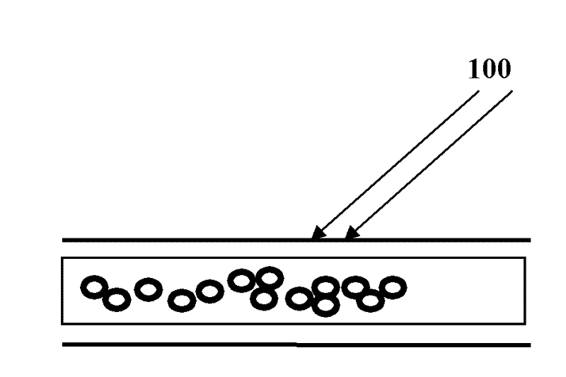
[0048] figure 1 Shown are 2D microfluidic chip cell detection results. Because the focus of the 2D microfluidic chip fluid can only be realized in the two-dimensional plane where the chip is located (that is, the sample liquid converges along the width direction under the sheath flow on the left and right sides), and the direction perpendicular to it is the depth of the channel. direction, convergence cannot be achieved. Therefore, it is possible that more than one cell molecule overlaps and passes through the detection point (where the laser beam is irradiated) at the same time, which affects the accuracy of the detection result.

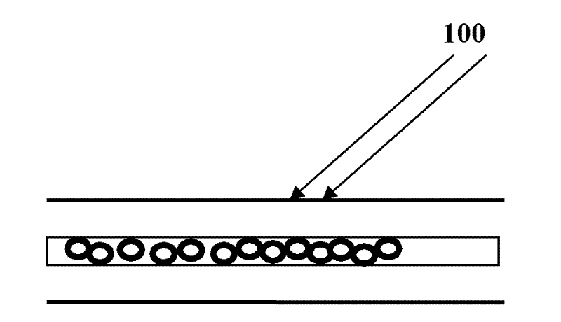
[0049] figure 2Shown is the cell detection result of the novel 3D microfluidic chip of the present invention. Due to the addition of a step structure in the converging channel, the fluid of the 3D microfluidic chip is also constrained in the vertical direction to achieve focus, so that only one cell passes through the detection point at the sam...


PUM


Abstract

The invention relates to a 3D (Three Dimensional) micro-fluidic structure for cell detection and a preparation method thereof. The entire structure is formed by connecting cover glass, a polydimethylsiloxane (PDMS) fluidic structure and a substrate in a sealing way, wherein a sheath fluid and sample fluid inflow channel (hole), a convergence cavity, a stepped structure and a convergence channel are carved on the PDMS fluidic structure, wherein the stepped structure and the convergence channel are used for realizing 3D focusing; in the convergence cavity, the designed channel shape can realize leftward and rightward focusing of fluid; and in the convergence channel, fluid flows through the stepped structure and is constrained in the vertical direction, so that the 3D focusing can be realized. Meanwhile, the invention provides a process method for preparing the micro-fluidic structure in a twice photoetching and one developing way. By adopting a simple structural scheme, 3D fluid focusing can be realized, and a good application effect and convenience for machining can be achieved; and the PDMS is taken as a structural material, so that low cost and high bio-compatibility are realized.

Description

technical field [0001] The invention relates to a structure type of a three-dimensional microfluidic chip used for flow cytometric detection and a preparation method thereof. Background technique [0002] Flow cytometry is a technique for automatic multi-parameter analysis and detection of cell particles in liquid flow one by one. It can more accurately realize the qualitative and quantitative analysis of cell characteristics and cell (or organelle) composition, so it has extremely important application value in modern medical research and clinical diagnosis. Flow cytometer (FCM) is a modern biomedical instrument based on the basic principles of flow cytometry, which comprehensively utilizes optics, mechanics, fluid dynamics and computer control technology to realize cell detection and analysis functions. One of its core components is a flow chamber system with a specific fluid control structure (channel); its basic function is realized by reasonably controlling the motion ...

Claims


Application Information

Patent Timeline
IPC IPC(8): G01N15/14
Inventor 王永泉陈花玲王小鹏王敬元李义平王冰
Owner XI AN JIAOTONG UNIV
Why Eureka
Try Eureka