CD106-positive cells, and identification and preparation method and application thereof

An identification method, a technology of positive cells, applied in the field of separation and preparation of CD106 mesenchymal stem cells

Active Publication Date: 2012-07-04
北京汉氏干细胞科技有限公司
View PDF6 Cites 13 Cited by
  • Summary
  • Abstract
  • Description
  • Claims
  • Application Information

AI Technical Summary

Problems solved by technology

Ren et al pointed out in the article published in the Journal of Immunology (Journal of Immunology, 2010; 184:2321-8) that inflammatory factors can upregulate ICAM-1 and VCAM-1 to enhance the immunosuppressive effect, and use IFN-γ to stimulate mesenchymal Stem cells increase VCAM-1 expression, but this also increases HLA-DR expression

Method used


Image

  • CD106-positive cells, and identification and preparation method and application thereof
  • CD106-positive cells, and identification and preparation method and application thereof
  • CD106-positive cells, and identification and preparation method and application thereof

Examples


Embodiment 1

[0024] Example 1: Isolation of placenta-derived cells and acquisition and expansion of CD106-positive cells.

[0025] 1. Placenta collection, the placenta was collected with the informed consent of the parturient. Wash the placenta with sterile normal saline, then put the placenta into a special aseptic collection box, and add the collection solution. The placentas were then transported to the experimental site at 4°C for no more than 48 hours.

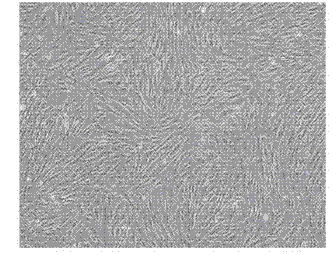
[0026] 2. Isolation of mesenchymal stem cells from placental chorionic tissue: (1) Separate the chorionic tissue by mechanical means; (2) Cut the tissue to 0.1-1 cubic centimeter, and then wash the tissue block repeatedly with phosphate solution5 (3) Add collagenase and trypsin mixture at a ratio of 1:1 to the tissue volume, and digest at a constant temperature of 37°C for 60 minutes; (4) Pass Use a 200-mesh filter to obtain a single-cell suspension, centrifuge, and remove the supernatant; (5) Resuspend the cells in DF12 medium.

...

Embodiment 2

[0034] Example 2: Sorting of CD106 positive mesenchymal stem cells.

[0035] The expanded mesenchymal stem cells were separated from the placenta-derived cells to separate CD106-positive cells by using a magnetic bead sorting method well known to professionals.

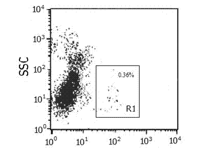
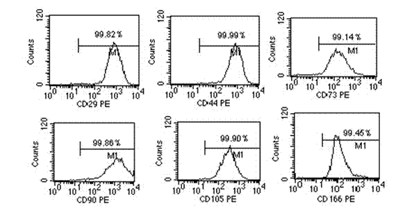
[0036] Results: After sorting by magnetic beads, the purity of CD106 positive cells obtained was 97.91%.

Embodiment 3

[0037] Example 3: CD106-positive mesenchymal stem cells express higher levels of COX2 / PGE2 and HGF.

[0038] The CD106-positive mesenchymal stem cells and CD106-negative mesenchymal stem cells were amplified to the required number respectively, and the culture supernatant was aspirated for future use, and the mRNA of the above two cells was extracted by a method well known to professionals, and reverse transcribed into cDNA. Quantitative polymerase chain reaction (rt-PCR) was used to compare the mRNA expression levels of COX2 and HGF in the two cells.

[0039] Use enzyme-linked immunosorbent assay (ELISA) well-known to professionals to detect the contents of PGE2 and HGF in the culture supernatant retained in the previous step.

[0040] Result: CD106 + mesenchymal stem cells than CD106 - Mesenchymal stem cells express higher levels of COX2 and HGF mRNA (see Figure 5 ), CD106 + mesenchymal stem cells than CD106 - Mesenchymal stem cells express higher levels of PGE2 and H...


PUM

PropertyMeasurementUnit
purityaaaaaaaaaa

Abstract

The invention discloses an identification and preparation method and application CD106-positive mesenchymal stem cells. The method comprises the following steps: identifying and isolating CD106-positive cells from mononuclear cells isolated from bone marrow, placenta or other tissues through an immunological method, carrying out adherent culture, and collecting adherent CD106-positive cells; or carrying out adherent culture of mononuclear cells isolated from bone marrow, placenta or other tissues, collecting adherent mesenchymal cells, and isolating the CD106-positive mesenchymal stem cells when the count of the obtained cells is large enough. The CD106-positive cells are mesenchymal stem cells with adherent growth, express mesenchymal stem cell-related immunological phenotype, and have osteogenesis, adipogenesis and other differentiation potencies. Compared with CD106-negative mesenchymal stem cells, the CD106-positive cells have higher immunosuppression capacity, can better inhibit proliferation of IFN-gamma (interferon-gamma) secretion of lymphocytes, and can be used for treating graft versus host disease (GVHD) and autoimmune diseases.

Description

technical field [0001] The invention relates to the separation and preparation of cells, and the patent relates to a method for the separation and preparation of CD106 mesenchymal stem cells with stronger immunosuppressive ability. Background technique [0002] A large number of in vivo and in vitro studies have shown that mesenchymal stem cells (MSCs) can directly inhibit the proliferation, differentiation and effector mechanisms of T cells, or indirectly by inhibiting the differentiation and maturation of dendritic cells (DCs). Inhibits the proliferation of T cells and affects the differentiation of effector T cell subsets. In addition to T lymphocytes and DCs, the target cells of MSCs also include many other types of immune cells, such as NK cells (natural killer cells, NK), B cells, neutrophils, macrophages, etc., which regulate the immune biology of these cells Activities, such as cytokine secretion, cytotoxic activity, etc., ultimately induce anti-inflammatory effects...

Claims


Application Information

Patent Timeline
Patent Type & Authority Applications(China)
IPC IPC(8): G01N33/53C12N5/0775C12N5/0735
Inventor 韩忠朝
Owner 北京汉氏干细胞科技有限公司
Why Eureka
Try Eureka