Methods for Ex Vivo Administration of Drugs to Grafts Using Polymeric Nanoparticles

a technology of polymer nanoparticles and nanoparticles, which is applied in the direction of prosthesis, surgery, catheters, etc., can solve the problems of autograft rejection, many patients experience life-threatening side effects, and autografts do not always adapt to life-threatening side effects, so as to facilitate the attachment of nanoparticles, inhibit hyperplasia, and prevent the rejection of grafts

Inactive Publication Date: 2010-06-17
YALE UNIV
View PDF54 Cites 29 Cited by
  • Summary
  • Abstract
  • Description
  • Claims
  • Application Information

AI Technical Summary

Benefits of technology

[0009]Methods for ex vivo administration of drugs to grafts using polymeric nanoparticles and applications for these methods are disclosed herein. These methods involve direct application of nanoparticles to graft materials prior to implantation in a host. The polymeric nanoparticles can have ligands which facilitate attachment of the nanoparticles to the graft and/or ligands which target the polymeric nanoparticles to specific cell or tissue types. Ligands may be directly attached to the polymer or may be attached to an adaptor element which associates with the polymeric nanoparticle. Ligands may be attached to adaptor elements directly via covalent bonds, or indirectly through an interaction between two molecules which form highly-specific, non-covalent physiochemical interactions. Molecules which prevent graft rejection or aid in the proper adaptation of the graft into the host can be encapsulated in or attached to...

Problems solved by technology

Still, allografts do not always survive and many patients experience life-threatening side effects from the powerful drugs that are used to prevent tissue rejection.
Autografts do not always adapt to their new location, and can in some cases adapt improperly, such as when venous bypasses in the coronary artery experience neointimal hyperplasia and stenosis.
Neointimal hyperplasia, the proliferation of vascular smooth muscle cells, is a major contributor to vessel restenosis and the principal cause of failure in late bypass grafts and arteriovenous graft fi...

Method used


Image

  • Methods for Ex Vivo Administration of Drugs to Grafts Using Polymeric Nanoparticles
  • Methods for Ex Vivo Administration of Drugs to Grafts Using Polymeric Nanoparticles
  • Methods for Ex Vivo Administration of Drugs to Grafts Using Polymeric Nanoparticles

Examples


example 1

Controlled Release of Drugs from Nanoparticles

[0096]Materials and Methods:

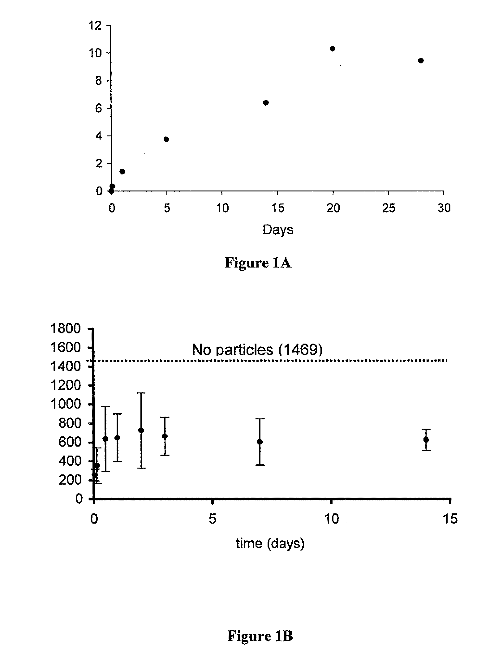
[0097]Five milligrams of rapamycin-loaded nanospheres were placed in 18 different tubes and suspended in 0.5 mls of phosphate buffered saline. The tubes were then incubated at 37° C. on a rotary shaker. At various time points, three of the tubes were removed and centrifuged to pellet the nanospheres. The supernatant was discarded and the nanospheres dissolved in NaOH to release all of the encapsulated rapamycin. The amount of released rapamycin was measured by absorption at 290 nm and converted to micrograms from a standard curve. This value, rapamycin remaining in the particles after PBS incubation, was then subtracted from the total amount of rapamycin encapsulated to yield the total amount released at that time point.

[0098]The bioactivity of the released rapamycin was then determined by PBMC assay. Briefly, PBMC cells were stimulated with IL-12 and IL-18. The levels of interferon released from PBMC cells we...

example 2

Localized Coating of Vascular Grafts with Nanoparticles

[0101]Materials and Methods:

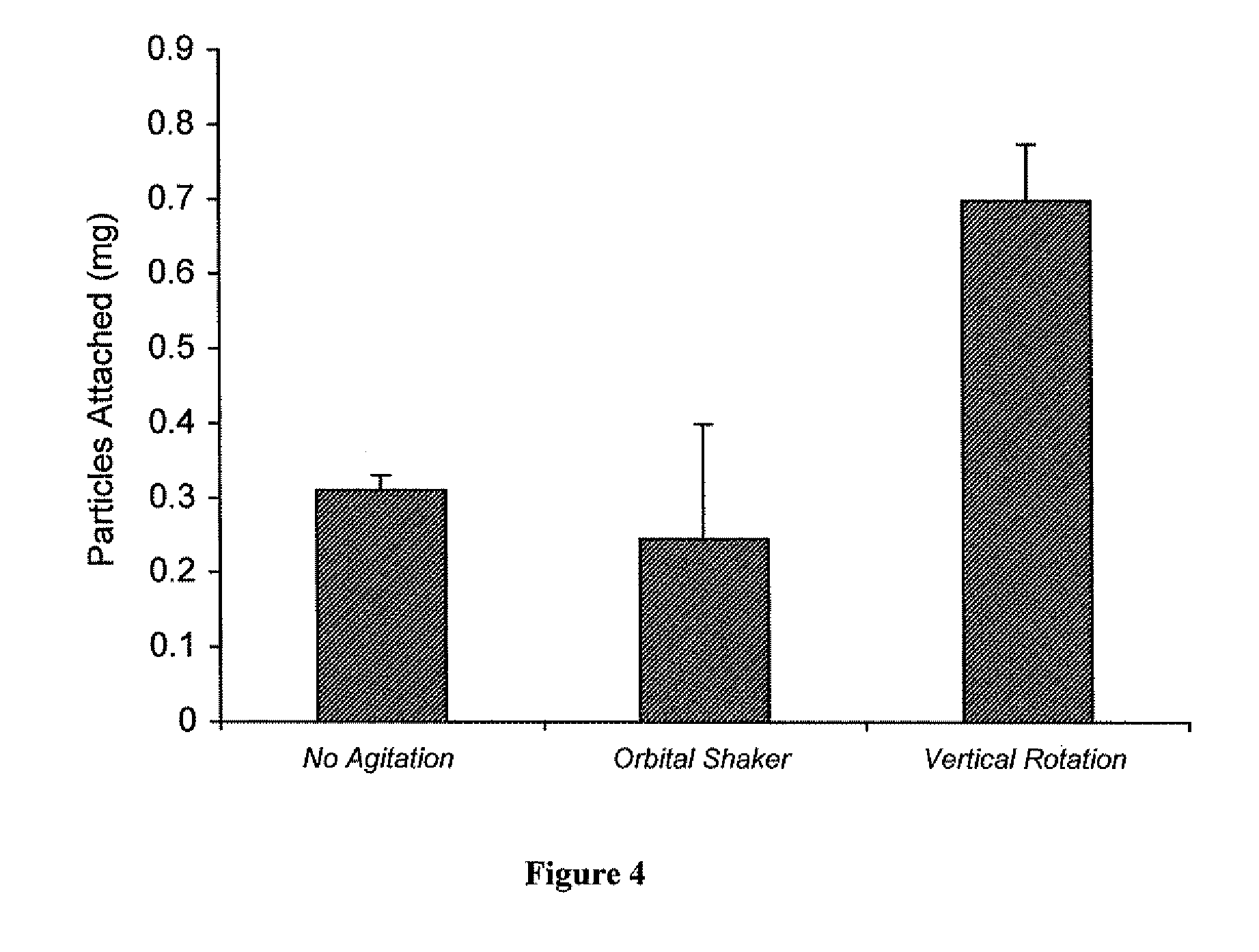
[0102]Rhodamine-loaded nanoparticles were used to demonstrate attachment (and impregnation) of nanoparticles to vascular wall of a human saphenous vein. Vascular grafts were either not coated, lumen- or intima-coated, adventia-coated, or both intima- and adventia-coated. To coat the vascular grafts 5 mgs of avidin-coated rhodamine nanoparticles were placed in a 5 ml scinillation vial and then suspended in a solution of Pluronic F-127 (300 μl of 10% Pluronic in DMSO and 2700 μl of PBS). The glass vial was then fixed to a vertical carousel of a hybridization oven and mixed for 20 minutes (maximal RPM, 25° C.). Parafilm was used to cover the portion of the vessel not to be coated.

[0103]Results:

[0104]Rhodamine nanoparticles were clearly visible in the intima of the graft which was intima-coated and likewise in the adventia of the graft which was adventia-coated. A greater degree of attached rhodamine nano...

example 3

Amount of Coating of Vascular Grafts with Nanoparticles

[0105]Materials and Methods:

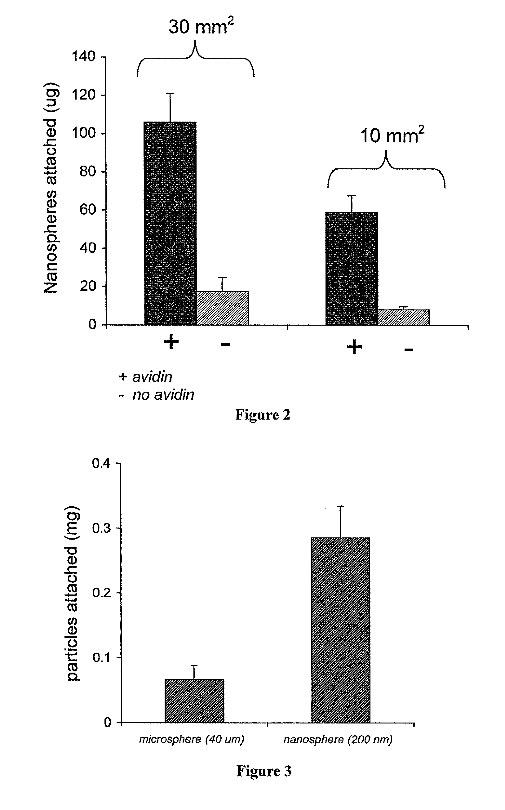
[0106]Two different sizes of ovine vascular tissue, 30 mm2 (n=3) and 10 mm2 (n=3), were coated with rhodamine nanoparticles and evaluated for amount of bound nanoparticles. The tissues were either coated with avidin rhodamine nanospheres or blank rhodamine nanospheres. After coating, the pieces of tissue were rinsed in distilled water three times, frozen and lyophilized. After two days the tissue was removed from the lyophilizer and suspended in 0.5 ml of DMSO for 4 hours to dissolve the nanospheres, thereby releasing the encapsulated rhodamine. The 0.5 ml of DMSO was then removed from the tissue and mixed with 0.5 ml of distilled water. This mixture was allowed to set for 30 minutes and then centrifuged to remove any precipitated polymer or loose tissue, and then scanned for fluorescence (ex. 550, em. 580). The resultant value was used to calculate the amount of particles attached.

[0107]Results:

[0108...


PUM

PropertyMeasurementUnit
Diameteraaaaaaaaaa
Diameteraaaaaaaaaa
Diameteraaaaaaaaaa

Abstract

Methods for ex vivo administration of drugs to grafts using polymeric micro- and nanoparticles and applications for these methods are described herein. The particles contain encapsulated molecules which are released locally at the site of implantation and function to prevent graft rejection or aid in the proper adaptation of the graft to the host. The disclosed methods may be used to inhibit or reduce hyperplasia and stenosis of vascular grafts or to prevent graft rejection.

Description

CROSS REFERENCE TO RELATED APPLICATIONS[0001]This application claims priority to and benefit of Provisional U.S. Patent Application No. 60 / 892,658 filed on Mar. 2, 2007.STATEMENT REGARDING FEDERALLY FUNDED RESEARCH[0002]This invention was made with government support awarded by the National Institutes of Health under Grant Nos. 1030899, 1A05838, 656001, NS45236, and DK070068. The United States Government has certain rights in this invention.FIELD OF THE INVENTION[0003]The present disclosure generally relates to the field of methods and nanoparticulate compositions for ex vivo administration of drugs to tissues and organs for transplantation.BACKGROUND OF THE INVENTION[0004]Tissue and organ transplantation is a major life-saving strategy, thanks to advances in surgical technique over the past 50 years. Recent progress in immunosuppression has also served to improve graft survival and patient health, making transplantation more common. Still, allografts do not always survive and many ...

Claims


Application Information

Patent Timeline
IPC IPC(8): A01N1/02
CPCA61L27/34A61L27/507A61L27/54A61L29/10A61L2400/12A61L31/10A61L31/16A61L2300/624A61L29/16
Inventor FONG, PETER M.SALTZMAN, WILLIAM MARKFAHMY, TAREK M.
Owner YALE UNIV
Why Eureka
Try Eureka