
Use of collection of plans to develop new optimization objectives

A planning and surface contouring technology, applied in the direction of radiological diagnostic instruments, applications, calculations, etc., can solve the problems of increasing fixed margin, cancer cannot be completely eradicated, etc., and achieve the effect of reducing radiation exposure

Active Publication Date: 2012-09-05
KONINKLIJKE PHILIPS ELECTRONICS NV
View PDF5 Cites 12 Cited by
  • Summary
  • Abstract
  • Description
  • Claims
  • Application Information

AI Technical Summary

Problems solved by technology

Because these organs are in such close proximity, the usual approach of adding a fixed margin to the target volume poses a risk to these sensitive organs
Without increasing the margin there is a risk that the cancer will not be completely eradicated

Method used


Image

  • Use of collection of plans to develop new optimization objectives
  • Use of collection of plans to develop new optimization objectives
  • Use of collection of plans to develop new optimization objectives

Examples


Embodiment Construction

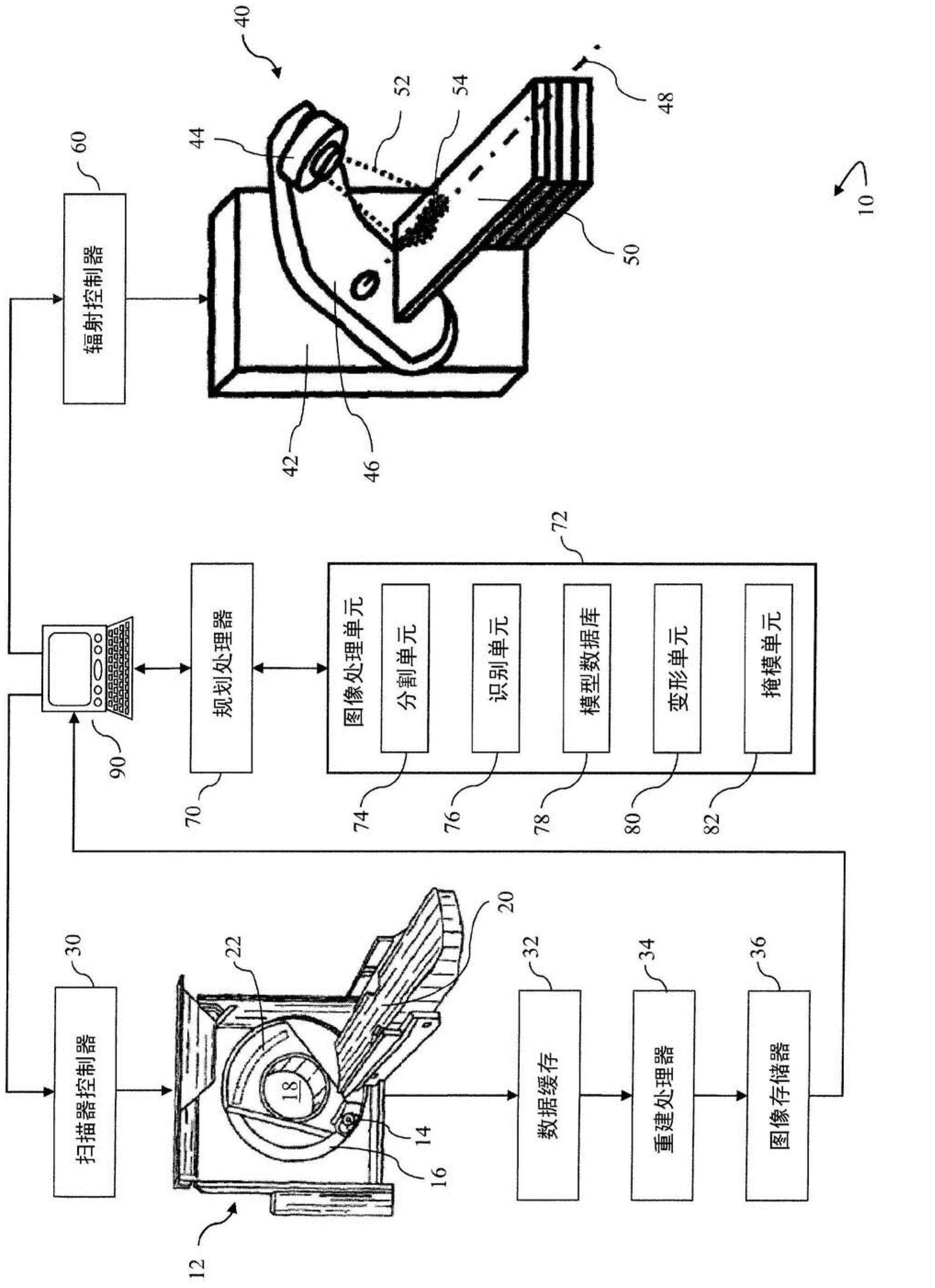
[0016] refer to figure 1 , a treatment system 10, such as a radiation therapy system, includes a diagnostic imaging scanner 12, such as a computed tomography (CT) imaging scanner, an MRI scanner, etc., for acquiring diagnostic images for use in planning radiation therapy protocols used in . CT imaging scanner 12 includes an x-ray source 14 mounted on a rotating gantry 16 . X-ray source 14 generates x-rays that pass through an examination region 18 where they interact with a target volume of a subject (not shown) supported by a support 20 that positions the target volume in the examination area within 18. The X-ray detector array 22 is arranged to receive the X-ray beam after it passes through the examination region 18 where it interacts with the subject and is partially absorbed by the subject. Accordingly, the detected X-rays include absorption information related to the subject.

[0017] CT scanner 12 is operated by controller 30 to perform a selected imaging sequence of...


PUM


Abstract

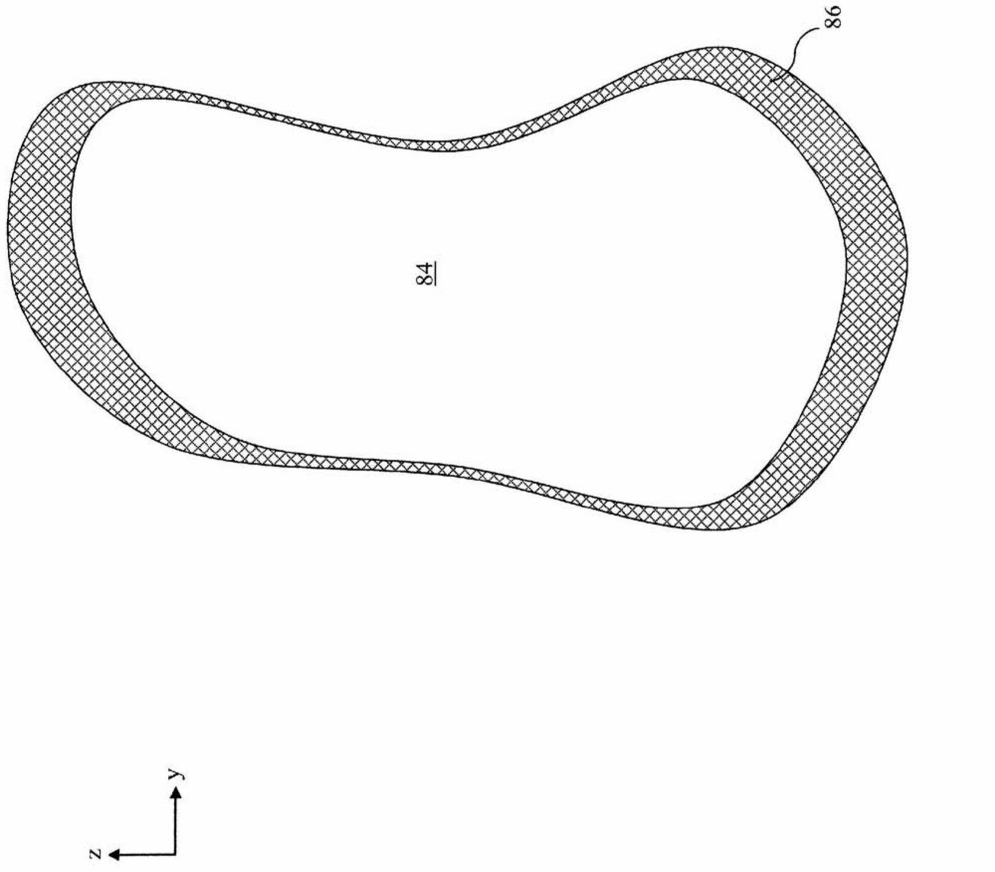
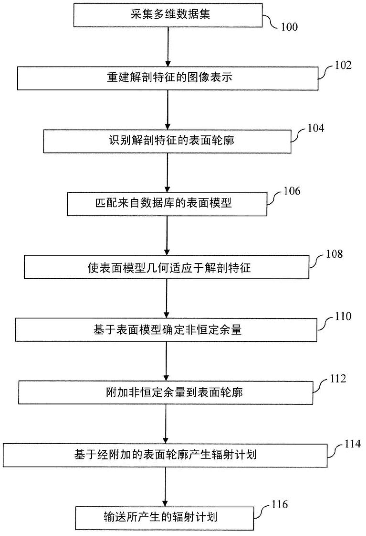
A radiation therapy system includes a diagnostic image scanner (12) which acquires a multidimensional dataset of a subject that is reconstructed into at least one image representation of an object of interest. An image processing apparatus (72), of radiation therapy system, includes a segmentation unit (74) which identifies a surface contour of the object of interest, or other critical structures. A masking unit (82) determines a non-constant margin, based on the identified surface contour and appends the determined non-constant margin to the identified surface contour. The non-constant margin is based on at least one of anisotropic motion, surface morphology, positional uncertainty, proximity to other organs, and probability of dose distribution. A planning processor (70) generates a radiation therapy plan which limits the delivery of therapeutic radiation to anatomy associated with the surface contour and appended non-constant margin. A radiation delivery system (40) delivers therapeutic radiation according to the generated plan.

Description

technical field [0001] The present application relates to methods and systems for radiation therapy planning. It finds particular application in identifying anatomical features in medical images and delivering radiation therapy to said anatomical features. Background technique [0002] Radiation therapy is a treatment technique commonly used in oncology in which doses of high-energy gamma (□) radiation, particle beams, or other radiation are delivered into a patient's body to achieve a therapeutic effect, namely eradication of cancerous tissue. Intensity modulation of multiple tomographic beams or a single beam using multi-leaf collimators or other beam shaping elements to precisely illuminate a target volume, e.g., cancerous tissue, while confining nearby sensitive organs at risk ) exposure. Radiation therapy session(s) are planned prior to radiation treatment based on one or more planning volume images, eg, computed tomography (CT) images, etc., of the tumor and its surr...

Claims


Application Information

Patent Timeline
IPC IPC(8): G06T7/00
CPCG06T2207/10072G06T2207/30096G06T7/12A61B5/0036A61B6/52A61B5/004A61N5/1039A61N5/103A61B6/5229A61B6/5211
Inventor M·巴尔
Owner KONINKLIJKE PHILIPS ELECTRONICS NV
Learn More