Supercharge Your Innovation With Domain-Expert AI Agents!

Image processing apparatus and x-ray diagnosis apparatus

A technology of image processing device and diagnostic device, which is applied in the fields of radiological diagnostic equipment, diagnosis, medical science, etc., and can solve problems such as complicated operation

Active Publication Date: 2012-11-28
TOSHIBA MEDICAL SYST CORP
View PDF12 Cites 8 Cited by
  • Summary
  • Abstract
  • Description
  • Claims
  • Application Information

AI Technical Summary

Problems solved by technology

However, there is a problem that the operation is complicated, or a difference is generated depending on the operator, and it is not desirable to operate in a clean area in endovascular treatment

Method used


Image

  • Image processing apparatus and x-ray diagnosis apparatus
  • Image processing apparatus and x-ray diagnosis apparatus
  • Image processing apparatus and x-ray diagnosis apparatus

Examples


Embodiment Construction

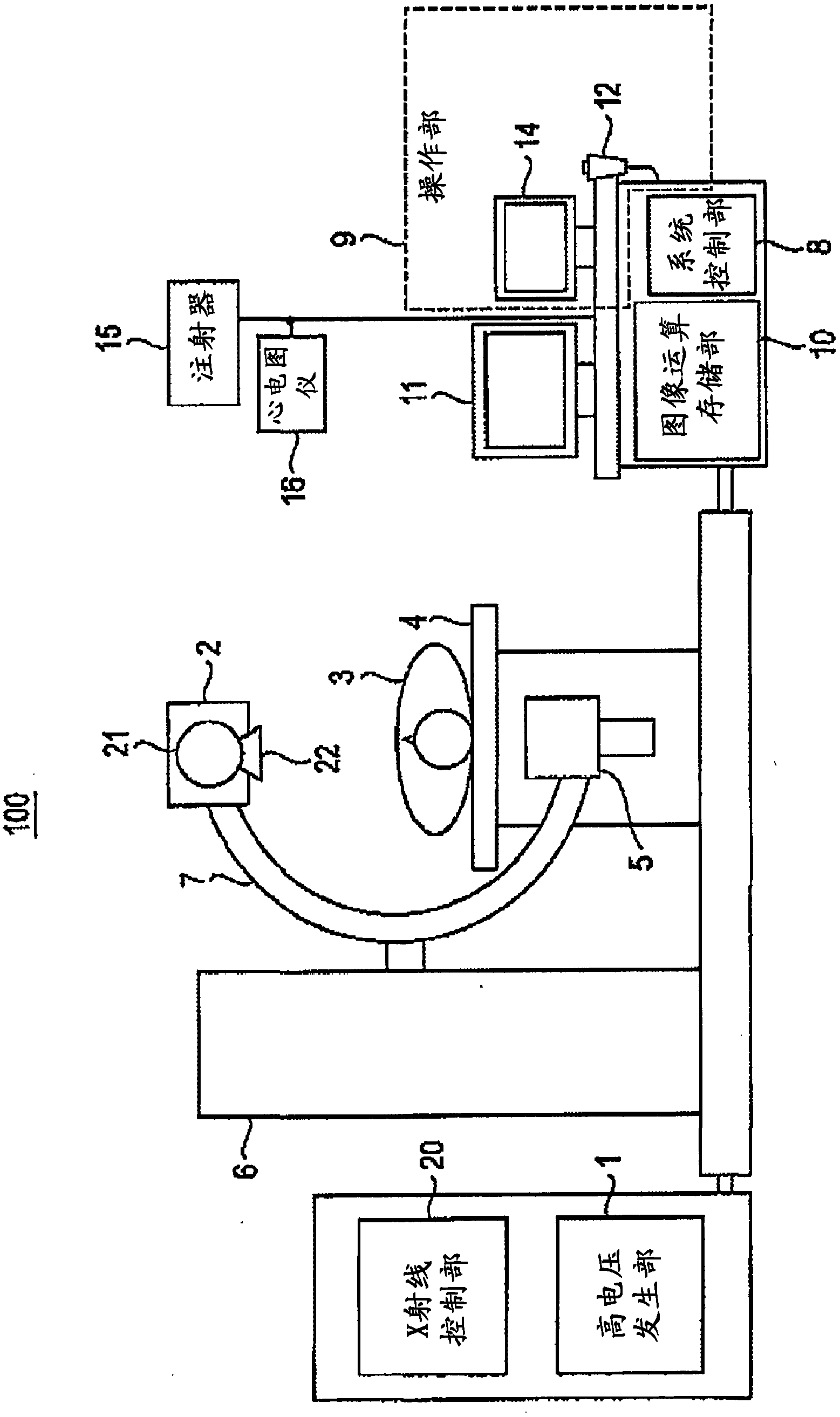
[0033] Hereinafter, an image processing device and an X-ray diagnostic device according to the present embodiment will be described with reference to the drawings.

[0034] figure 1 The X-ray diagnostic apparatus according to this embodiment is shown. The X-ray diagnostic apparatus has a gantry 100 . The frame 100 has a C-arm 7 . The C-arm 7 is rotatably supported by the support mechanism 6 . The X-ray generator 2 is attached to one end of the C-arm 7 . The X-ray generator 2 has an X-ray tube 21 and an X-ray collimator 22 . The high voltage generator 1 generates a high voltage (tube voltage) applied between electrodes of the X-ray tube 21 and also generates a filament current supplied to a filament of the X-ray tube 21 . The X-ray control unit 20 controls the tube voltage and the filament current generated by the high voltage generating unit 1 in accordance with the control of the system control unit 8 .

[0035] The X-ray detection unit 5 is attached to the other end of...


PUM


Abstract

The invention provides an image processing apparatus and an X-ray diagnosis apparatus, which enables the automatic setting of region of interest to be realized when a catheter operation is carried out. An image operating / storage unit (10) stores first X-ray image data in which a blood vessel of an object is not visually enhanced and second X-ray image data in which the blood vessel is visually enhanced via a contrast agent put to the detected object, specifies a catheter image included in the first fluoroscopic image by performing image processing of the first X-ray image data, extracts a blood vessel region included in the second fluoroscopic image by performing image processing of the second X-ray image data, and sets a region of interest based on the position of end point of the catheter.

Description

technical field [0001] Embodiments of the present invention relate to an image processing device and an X-ray diagnostic device. Background technique [0002] An X-ray image processing device for a circulator is being developed as an image guidance device for angiography or interventional treatment targeting arteries and veins of the whole body. Here, the X-ray image processing device processes an image captured by X-rays in a state in which a contrast medium is injected into a blood vessel, and displays the image data on an image display unit or an image display device. [0003] For example, in a coronary angiogram of the heart, a contrast agent is injected through an inserted catheter into the entrance of the coronary arteries. In addition, as an arterial examination, the state of stained blood vessels was measured, and as a histological examination, the state of stained myocardial tissue was measured. Among the latter, there is known a method of measuring blood flow in ...

Claims


Application Information

Patent Timeline
IPC IPC(8): A61B6/00
Inventor 姚敬武坂口卓弥
Owner TOSHIBA MEDICAL SYST CORP
Features
  • R&D
  • Intellectual Property
  • Life Sciences
  • Materials
  • Tech Scout
  • Unparalleled Data Quality
  • Higher Quality Content
  • 60% Fewer Hallucinations
Learn More