Eureka AIR delivers breakthrough ideas for toughest innovation challenges, trusted by R&D personnel around the world.

Automatic dividing method of ultrasound carotid artery plaque

An automatic segmentation and carotid artery technology, applied in ultrasound/sonic/infrasonic Permian technology, ultrasound/sonic/infrasonic image/data processing, image analysis, etc., can solve problems such as inability to obtain results

Active Publication Date: 2012-11-28
HUAZHONG UNIV OF SCI & TECH
View PDF3 Cites 33 Cited by
  • Summary
  • Abstract
  • Description
  • Claims
  • Application Information

AI Technical Summary

Problems solved by technology

The above two articles, after the contour of the inner and outer membranes are obtained by segmentation, shrink it inward for a certain distance as the initial contour, and then evolve to obtain accurate segmentation results, but both only consider gray-scale features for the segmentation of mixed plaques, which has limitations sex, the desired result cannot be obtained
All the segmentation methods mentioned above are for MRI images, CT images and intravascular ultrasound images. So far, there is no report on plaque segmentation methods in B-ultrasound carotid artery images

Method used


Image

  • Automatic dividing method of ultrasound carotid artery plaque
  • Automatic dividing method of ultrasound carotid artery plaque
  • Automatic dividing method of ultrasound carotid artery plaque

Examples


Embodiment Construction

[0087] The present invention will be further described in detail below in conjunction with the accompanying drawings and embodiments.

[0088] The plaque automatic segmentation algorithm of plaque in the ultrasonic image provided by the present invention, its implementation steps are as follows:

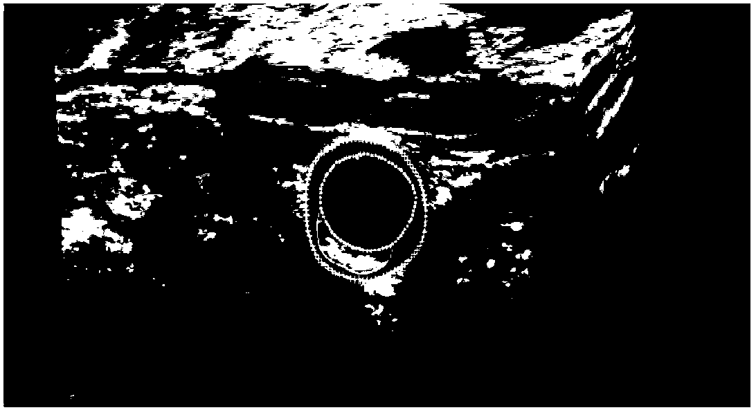
[0089] 1 Extract the current frame image from the three-dimensional ultrasound volume data of the carotid artery, and read the resolution ρ of the unit pixel in the horizontal and vertical directions corresponding to the image x and ρ y , ρ x is the number of pixels corresponding to 1mm in the horizontal direction, ρ y is the number of pixels corresponding to 1 mm in the vertical direction.

[0090] 2 Segment the current frame image to obtain the inner and outer membrane contours.

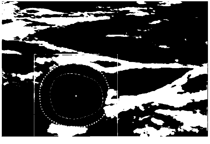
[0091] 3. Extract the region of interest (ROI) for plaque segmentation according to the contour of the inner and outer membranes, so as to reduce the computational overhead of the segmentation steps d...


PUM


Abstract

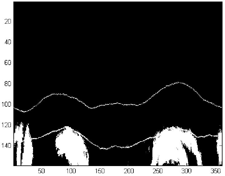
The invention belongs to the field of intersection of a computer technology and medical images, and in particular relates to a dividing method of plaque in the cross section direction of carotid artery blood vessel in an ultrasound image. The method specifically comprises the following steps of: selecting the current frame image; dividing to obtain profiles of internal and external membranes of the blood vessel; extracting the plaque and dividing the interested area; detecting an initial plaque external boundary: carrying out curve fitting on each column of pixel point gray value between the internal and external membrane profiles on a polar coordinate image obtained through conversion in the interested area, and detecting the pixel point with the minimum gray value and nearest to the external membrane profile as the initial plaque external boundary point; carrying out level set evolution to obtain the final plaque external boundary; and using the area between the final external boundary and the internal membrane profile as the plaque area. By adopting the dividing method of ultrasound carotid artery plaque, plaque can be accurately divided, the workload of doctors can be greatly reduced, and the plaque size and area calculated on the basis of the division result can help doctors analyze the lesion degree and treatment effect.

Description

technical field [0001] The invention belongs to the intersecting field of computer technology and medical images, and in particular relates to an automatic segmentation method for plaques in the cross-sectional direction of a carotid artery in an ultrasonic image. Background technique [0002] According to the statistics of the World Health Organization, the number of deaths caused by cardiovascular diseases accounts for about one-third of the total number of deaths in the world every year, and carotid atherosclerosis is closely related to cardiovascular and cerebrovascular diseases. Compared with the traditional Intima-Media Thickness (IMT) measurement index, the total plaque volume (Total Plaque Volume, TPV), plaque area (Total Plaque Area, TPA) and plaque composition analysis are more accurate. It can accurately and intuitively reflect the status of atherosclerosis, and has become one of the important indicators for predicting the risk of cardiovascular and cerebrovascula...

Claims


Application Information

Patent Timeline
Patent Type & Authority Applications(China)
IPC IPC(8): G06T7/00A61B8/08G06T7/11
Inventor 丁明跃程洁玉李鹤杨鑫
Owner HUAZHONG UNIV OF SCI & TECH
Why Eureka
Eureka Blog
Learn More