
Optic disc projection location method synthesizing vascular distribution with video disc appearance characteristics

A technology of appearance characteristics and positioning methods, used in image analysis, image data processing, instrumentation, etc.

Inactive Publication Date: 2012-12-26
XIANGTAN UNIV
View PDF2 Cites 43 Cited by
  • Summary
  • Abstract
  • Description
  • Claims
  • Application Information

AI Technical Summary

Problems solved by technology

[0008] In order to solve the above-mentioned technical problems existing in the existing optic disc positioning, the present invention provides an optic disc projection positioning method that integrates blood vessel distribution and appearance characteristics

Method used


Image

  • Optic disc projection location method synthesizing vascular distribution with video disc appearance characteristics
  • Optic disc projection location method synthesizing vascular distribution with video disc appearance characteristics
  • Optic disc projection location method synthesizing vascular distribution with video disc appearance characteristics

Examples


Embodiment Construction

[0068] The present invention will be described in further detail below in conjunction with the accompanying drawings and embodiments.

[0069] like figure 1 As shown, the optic disc projection positioning method of the present invention, its specific process is:

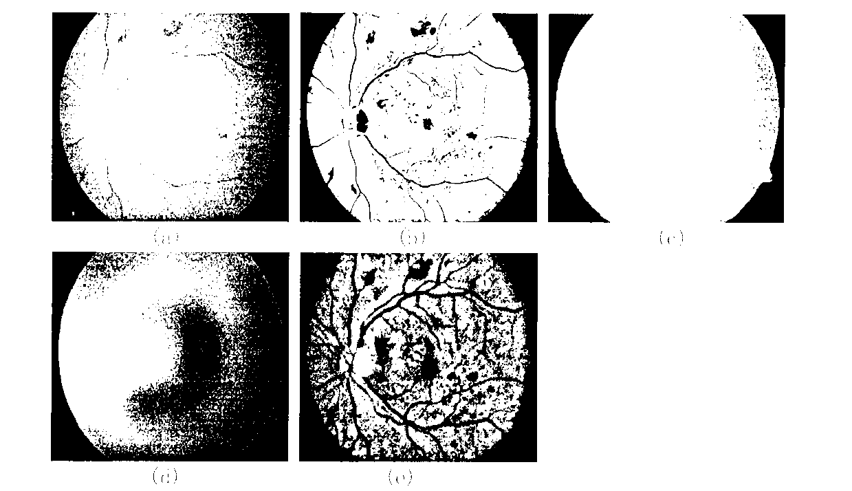
[0070] 1. Fundus image mask processing. like figure 2 As shown in (a), the retinal fundus image usually contains a dark background and the retinal fundus imaging area, where the retinal fundus imaging area is the area of ​​interest of the ophthalmologist. In order to prevent the interference of the background and the border, it is necessary to obtain the mask template of the area of ​​interest . Since the red component of the color fundus image is close to saturation and can best reflect the lighting conditions during imaging, the red channel component I in the original color fundus image is taken R 5% of the brightest pixel intensity value is used as the segmentation threshold t b , and segment the binary imag...


PUM


Abstract

The invention discloses an optic disc projection location method synthesizing vascular distribution with video disc appearance characteristics, which comprises the following steps of: (1) extracting an interest retina fundus image region by use of a mask operation; (2) carrying out normalization enhancement on a fundus image based on an image observation model; (3) achieving extraction and segmentation of the fundus image by using a non-vascular structure inhibition operator and combining with a hysteresis multi-threshold processing technology; (4) setting a vertical window of double main blood vessel width to slide on a blood vessel segmentation image along the horizontal direction and calculating a vascular distribution degree D(x) at each horizontal position x to obtain a distribution degree curve of a horizontal projection, wherein the minimum value point of the curve is determined to be optic disc horizontal coordinate xod; (5) setting a rectangular window side length of which is equal to the optic disc diameter to slide up and down at the horizontal coordinate xod, estimating local region brightness IN(xod, y) and edge gradient information gN(xod, y) respectively, drawing a vertical projection curve reflecting a change of a characteristic value f(y)=IN(xod, y)*gN(xod, y), wherein a maximum value point of the curve is optic disc vertical coordinate yod. The method has a simple algorithm, a high success rate and excellent robustness.

Description

technical field [0001] The invention relates to a method for automatically locating an optic disc in a retinal fundus image, in particular to an optic disc projection positioning method that integrates the distribution of blood vessels and the appearance characteristics of the optic disc. Background technique [0002] The optic disc is one of the main physiological structures of the retina. The optic nerve and blood vessels enter the eye from this area and radiate to the periphery. In the retinal fundus image, it appears as a round bright yellow area and contains a large number of thick blood vessels. Researchers have always paid great attention to the automatic positioning of the optic disc, because the accurate positioning of the optic disc is beneficial to the diagnosis of fundus lesions, for example: many optic disc segmentation algorithms need to give the initial seed point in advance; The distance is approximately constant, and when the location of the optic disc is de...

Claims


Application Information

Patent Timeline
IPC IPC(8): G06T7/00G06T5/00
Inventor 张东波易瑶王俊超尚星宇李雄
Owner XIANGTAN UNIV
Learn More