Method for detecting human amniotic epithelial cell molecular mechanism for in vitro immune regulation of lymphocyte

A technology of amniotic membrane epithelial cells and molecular mechanisms, applied in the field of detection of molecular mechanisms of human AECS in vitro immunosuppression

Inactive Publication Date: 2013-04-17
吴卫江
View PDF0 Cites 1 Cited by
  • Summary
  • Abstract
  • Description
  • Claims
  • Application Information

AI Technical Summary

Problems solved by technology

[0003] Previous experiments have verified that AECS has a low expression of HLA-DR and has an inhibitory effect on activated T cells, but whether AECS expresses co-stimulatory molecules related to T cell activation or inhibition and the mechanism involved has not been reported in the literature

Method used


Image

  • Method for detecting human amniotic epithelial cell molecular mechanism for in vitro immune regulation of lymphocyte
  • Method for detecting human amniotic epithelial cell molecular mechanism for in vitro immune regulation of lymphocyte
  • Method for detecting human amniotic epithelial cell molecular mechanism for in vitro immune regulation of lymphocyte

Examples


Embodiment Construction

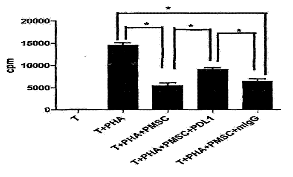
[0023] A detection method of the molecular mechanism of human AECS in vitro immunosuppressive action of the present invention, the research purpose of which is to further understand the immune regulation ability of human amnion cells and its possible effect mechanism, so as to provide the final clinical application of this technology and expand its clinical application indications Provide a theoretical basis.

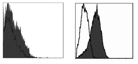
[0024] The materials and equipment used in this experiment include an inverted phase contrast microscope (Olympus, Japan), a C02 incubator (Jouan, France), a constant temperature centrifuge (Jouan, France), a flow cytometer (Beckman. Coulter, USA), culture flasks and Culture plate (Cotingcostar, USA), β liquid scintillation counter (Pharmac ia, Uppsala, Sweden), LG-DMEM medium (Hyclone), fetal bovine serum (Hyclone), trypsin (Sigma), DMEM (Gibco company), mouse anti-human PD1, CD80, CD83, CD86, CD4, CD69 cloned antibody (eBioscienee company), mouse anti-human B7H3mAb, P...


PUM

PropertyMeasurementUnit
purityaaaaaaaaaa

Abstract

The invention provides a method for detecting a human placenta-originated mesenchymal stem cells (MSCs) mechanism with the effects of inhibiting the activity and the proliferation of T cells in vitro. In view of the important role of costimulatory molecules in the activation and the proliferation of the T cells, the invention provides a bold speculation that the human placenta-originated MSCs show a negative regulation effect on the T cells in vitro, which may be related to the high-expression negative costimulatory molecules namely programmed death ligand-1 (PD-L1). The method comprises the steps of: firstly verifying that PMSCS (Placentaderived Mesenchymal Stem Cells) has an obvious inhibiting effect on the T-cell activation through experiments, taking negative costimulatory molecules namely the PD-L1 as an entry point, analyzing the role of the PD-L1 in the PMSCS-mediated effects of inhibiting the activity and the proliferation of the T cells in vitro through experiments such as flow cytometer detection, immunofluorescent staining, 3H-TdR (3H-Thymidine Riboside) mixing method, ELISA (Enzyme Linked Immunosorbent Assay) and the like and accordingly verifying that the PD1-PD-L1 molecular pathway is the main signaling mechanism of the PMCSC for regulating the immune tolerance of the T cells in vitro.

Description

technical field [0001] The invention relates to the fields of biotechnology and immunology, in particular to a detection method for the molecular mechanism of human AECS immunosuppression in vitro. Background technique [0002] Co-stimulatory molecules play an important role in the activation and proliferation of T lymphocytes. "Co-stimulatory signal" was proposed by Bretseher and Cohn in 1970 based on the dual-signal model of T cell activity, that is, in addition to presenting the MHC-treated antigen through APC to provide the first signal to antigen-specific T cells, it also Co-stimulatory molecules are required to act as auxiliary signals, and T cells can produce normal immune regulation mechanisms after reaching the physiological activation threshold. The absence of a secondary signal from costimulatory molecules will lead to anergy or specific immune tolerance of regulatory T cells. The B7 family is the only co-stimulatory molecule that can transmit signals from APC t...

Claims


Application Information

Patent Timeline
Patent Type & Authority Applications(China)
IPC IPC(8): C12Q1/02G01N33/68
Inventor 徐杰房文峰朱爱华
Owner 吴卫江
Why Eureka
Try Eureka