

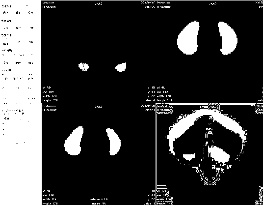
Quantitative analysis software for pulmonary perfusion and ventilation tomography
A technology of tomographic imaging and image analysis, applied in special data processing applications, instruments, electrical digital data processing, etc., can solve the difficult problems of quantitative analysis and quantitative curative effect evaluation of pulmonary embolism
- Summary
- Abstract
- Description
- Claims
- Application Information
AI Technical Summary
Problems solved by technology
Method used
Image


Examples
Embodiment Construction
[0008] one. software function
[0009] 1. image registration
[0010] The image of the patient can be registered and fused through the software, and the software provides two registration methods: automatic registration and manual registration. Automatic registration takes a period of time, and the system automatically registers according to the image data and automatically displays the registered and fused image in the specified view display area; manual registration is the way the user uses his own manual operation to manually move the image Complete the registration of the two views according to your own visual judgment, and then click the Finish button to end the manual registration operation.
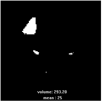
[0011] 2. analysis editor
[0012] It is analyzed and rendered into two-dimensional or three-dimensional images using the functions of the control system. To a certain extent, doctors can use the software to perform some auxiliary operations on the patient's condition analysi...
PUM
 Abstract
Description
Claims
Application Information