
Ultrasonic imaging method and ultrasonic imaging device

A technology of ultrasonic imaging and ultrasonic image, applied in the directions of ultrasonic/sonic/infrasonic image/data processing, ultrasonic/sonic/infrasonic diagnosis, ultrasonic/sonic/infrasonic Permian technology, etc., which can solve the problem of low contrast resolution, It is impossible to clearly show the perfusion of contrast medium and affect the differential diagnosis, so as to avoid the effect of controlling the process

Active Publication Date: 2013-06-05
SHENZHEN MINDRAY BIO MEDICAL ELECTRONICS CO LTD +1
View PDF4 Cites 13 Cited by
  • Summary
  • Abstract
  • Description
  • Claims
  • Application Information

AI Technical Summary

Problems solved by technology

In the ultrasonic echo reflected by human tissue containing contrast agent, the linear component includes both the linear component of the tissue and the linear component of the contrast agent. The contrast resolution of the ultrasonic image formed by the detection and processing of the linear component of the fundamental wave is not high, and the contrast cannot be clearly presented. The perfusion of agents in microvessels and tissues affects clinical differential diagnosis

Method used


Image

  • Ultrasonic imaging method and ultrasonic imaging device
  • Ultrasonic imaging method and ultrasonic imaging device
  • Ultrasonic imaging method and ultrasonic imaging device

Examples


Embodiment Construction

[0023] Such as figure 1 As shown, the device for ultrasonic imaging of the target area according to the embodiment of the present invention includes: a probe 1, a transmitting circuit 2, a transmitting / receiving selection switch 3, a receiving circuit 4, a beam forming module 5, a signal processing module 6, and an image processing module 7 and display 8.

[0024] The transmitting circuit 2 transmits the delayed-focused ultrasonic pulse with a certain amplitude and polarity to the probe 1 through the transmitting / receiving selection switch 3 . Probe 1 is excited by ultrasonic pulses, and emits ultrasonic waves to the target area (not shown in the figure) of the body tissue under test, and receives the ultrasonic echoes with tissue information reflected from the target area after a certain delay, and sends the The ultrasound echoes are converted back into electrical signals. The receiving circuit receives the electrical signal converted and generated by the probe 1 to obtain ...


PUM


Abstract

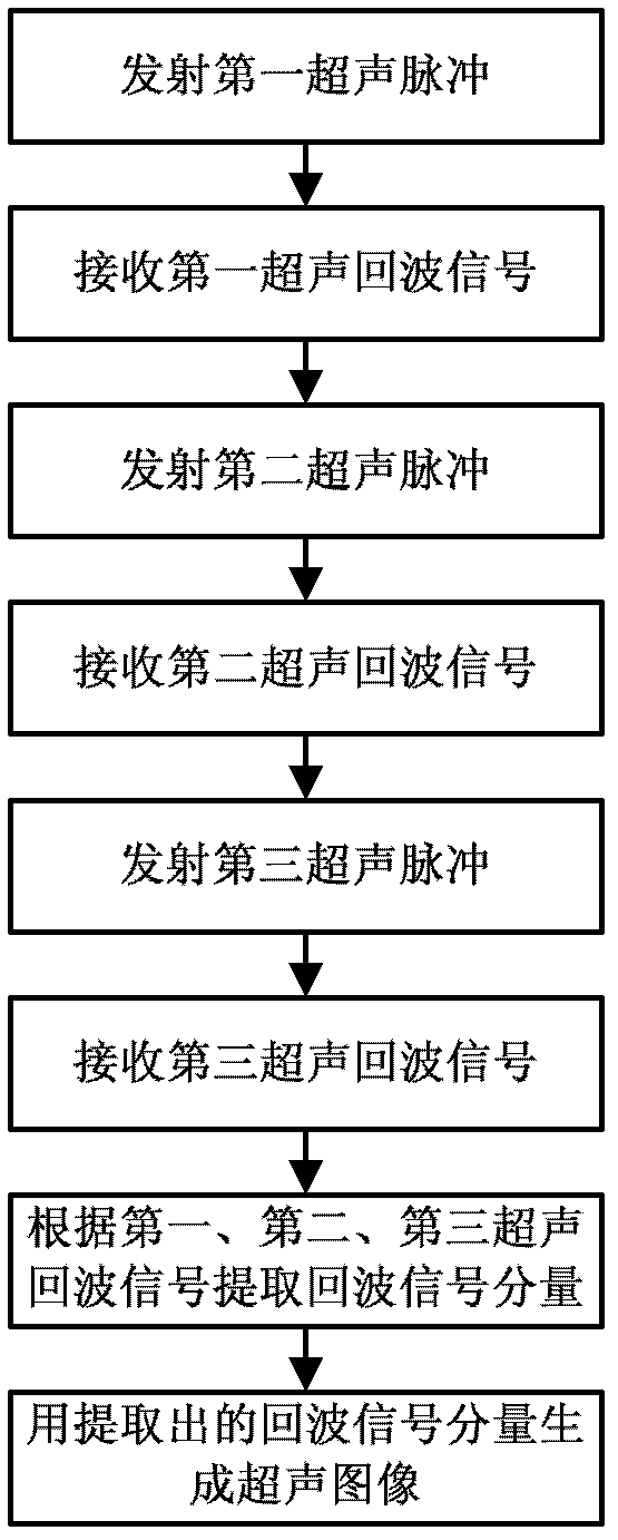
The invention discloses a method and a device for performing ultrasonic imaging on a target area. The ultrasonic imaging method includes sending a first ultrasonic pulse and receiving a first ultrasonic echo signal, sending a second ultrasonic pulse and receiving a second ultrasonic echo signal, sending a third ultrasonic pulse and receiving a third ultrasonic echo signal, extracting an echo signal component according to the first ultrasonic echo signal, the second ultrasonic echo signal and the third ultrasonic echo signal, and generating an ultrasonic image of the target area according to the echo signal component, wherein the amplitude weight of the third ultrasonic pulse is equal to the sum of the amplitude weight of the first ultrasonic pulse and the amplitude weight of the second ultrasonic pulse. The plurality of ultrasonic echo signals are processed and modulated, asymmetric components of even order nonlinear components and odd order nonlinear components in the ultrasonic echo signals are enabled to be separated from symmetric components of linear components and the odd order nonlinear components, and the asymmetric components and / or the even order nonlinear components of the odd order nonlinear components can be conveniently extracted from the ultrasonic echo signals.

Description

technical field [0001] The invention relates to the field of medical ultrasonic imaging, in particular to a method and device for ultrasonic imaging of a target area. Background technique [0002] In the medical ultrasound imaging system, the transmitting circuit usually transmits ultrasonic pulses into the human body, and the visible ultrasonic images of human tissues are obtained by receiving and processing the echoes carrying the characteristic information of human tissues by using the reflection of ultrasonic waves at the interface of human tissues. [0003] In ultrasound imaging, due to the existence of reverberation and the limitation of resolution, ultrasound images sometimes appear blurred or even unable to display weak boundaries and small blood vessels. In contrast agent imaging, the acoustic impedance difference between the contrast agent and the surrounding tissue is large, which can change the absorption, reflection, scattering and refraction of sound waves betw...

Claims


Application Information

Patent Timeline
IPC IPC(8): A61B8/08A61B8/00
CPCG01S7/52038G01S7/5202
Inventor 桑茂栋冒祖华吉挺澜
Owner SHENZHEN MINDRAY BIO MEDICAL ELECTRONICS CO LTD
Learn More