
A Sparse Angle X-ray CT Imaging Method

A sparse-angle, CT imaging technology, applied in image data processing, 2D image generation, instruments, etc., can solve the problems of inability to accurately reflect the characteristics of imaging organs, unfavorable and accurate judgment, etc.

Inactive Publication Date: 2016-05-18
SOUTHERN MEDICAL UNIVERSITY
View PDF3 Cites 0 Cited by
  • Summary
  • Abstract
  • Description
  • Claims
  • Application Information

AI Technical Summary

Problems solved by technology

The PICCS method directly reconstructs the current image without processing the previously scanned CT image through the constructed objective function, so that there are obvious artifacts in places where the imaging positions are inconsistent, and if only a simple registration method is used to eliminate the artifacts, Imaging results cannot accurately reflect the characteristics of the imaging organs, which is not conducive to making accurate judgments clinically

Method used


Image

  • A Sparse Angle X-ray CT Imaging Method
  • A Sparse Angle X-ray CT Imaging Method
  • A Sparse Angle X-ray CT Imaging Method

Examples


Embodiment 1

[0044] A sparse-angle X-ray CT imaging method includes the following steps in sequence.

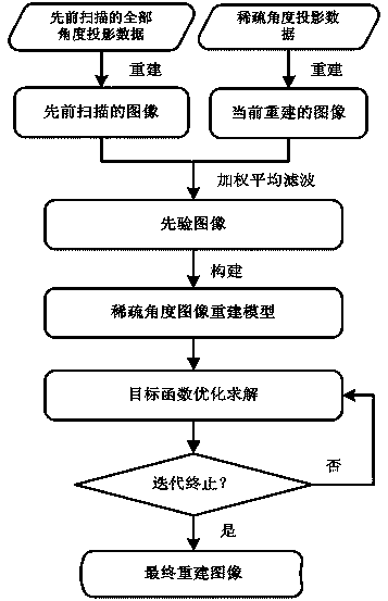
[0045] (1) Obtain CT system parameters, all angle projection data from previous scans, and sparse angle projection data at different time periods.

[0046] (2) Reconstruct images using the CT reconstruction method for all angle projection data and sparse angle projection data obtained in step (1) of the previous scan to obtain the CT image of the previous scan and the current reconstructed CT image .

[0047] Wherein, the CT reconstruction method may be a filtered back projection method or an iterative reconstruction method, or other methods known in the art.

[0048] (3), CT image obtained by step (2) previously scanned and the current reconstructed CT image , using weighted average filtering to obtain the prior image for sparse angle CT image reconstruction .

[0049] The weighted average filtering process adopted above is specifically carried out by the following formula:

...

Embodiment 2

[0068] In this embodiment, the specific implementation process of the reconstruction method of the present invention is described in detail by taking the sparse angle CT image obtained by the revised Shepp-Logan phantom simulation as an example.

[0069] Such as figure 1 As shown, the implementation process of this embodiment is as follows.

[0070] Step (1), set the parameters of the CT imaging geometric system, and obtain the system matrix , the sampling value of the projection angle in one week is 1160 and is equally spaced sampling, each projection angle corresponds to 672 detector units, and the size of the detector unit is 1.407mm.

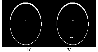
[0071] According to the standard Shepp-Logan phantom ( figure 2 (a) The simulation obtains 1160 projection data of all angles .

[0072] The standard Shepp-Logan phantom is revised, adding two motion areas, and then setting the sampling value of the projection angle within one week of scanning to 25 and sampling at equal intervals. Ac...


PUM


Abstract

A sparse angle X-ray captive test (CT) imaging method comprises the following steps: (1) system parameters, all angle projection data scanned previously and sparse angle projection data scanned previously in different periods are obtained; (2) image reconstruction is respectively performed to the all angle projection data scanned previously and the sparse angle projection data scanned previously which are obtained in step (1) so that a previously scanned CT image and a currently reconstructed CT image are obtained; (3) weighting even filtering processing is performed to the previously scanned CT image and the currently reconstructed CT image which are obtained in the step (2) so that a prior image is obtained; (4) a sparse angle CT image reconstruction model is constructed by the prior image obtained in the step (3); and (5) a final reconstructed image is obtained through optimization solution. According to the sparse angle X-ray CT imaging method, motion artifact caused by mismatching of imaging positions between the previously scanned CT image and the currently reconstructed CT image is removed so that high quality reconstruction of a low dose CT image is achieved.

Description

technical field [0001] The invention relates to the technical field of image processing of medical images, in particular to a sparse-angle X-ray CT imaging method. Background technique [0002] X-ray CT scanning has been widely used in clinical medical imaging diagnosis, but excessive X-ray radiation dose during CT scanning may cause cancer risk. In order to reduce the damage to users, how to minimize the dose of X-rays has become one of the key technologies in the field of medical CT imaging. [0003] In order to reduce the dose of X-ray radiation, various hardware and software technologies can be used to reduce the dose of X-ray used in CT scanning. Common methods include reducing tube current, reducing X-ray exposure time, and reducing projection data. The reduction of projection data is sparse-angle CT scanning. [0004] Due to missing projection data in sparse-angle CT scans, image reconstruction is required to obtain a good image. For sparse angle reconstruction, du...

Claims


Application Information

Patent Timeline
Patent Type & Authority Patents(China)
IPC IPC(8): G06T11/00
Inventor 马建华张蕴婉黄静曾栋高杨陈武凡
Owner SOUTHERN MEDICAL UNIVERSITY
Learn More