Method for determining cancer onset or cancer onset risk

A determination method, cancer technology, applied in disease diagnosis, material excitation analysis, nanotechnology for sensing, etc., can solve the problem of low detection sensitivity

Inactive Publication Date: 2013-06-19
TOHOKU UNIV
View PDF7 Cites 3 Cited by
  • Summary
  • Abstract
  • Description
  • Claims
  • Application Information

AI Technical Summary

Problems solved by technology

Therefore, the detection sensitivity is low, and there are also quantitative problems

Method used


Image

  • Method for determining cancer onset or cancer onset risk
  • Method for determining cancer onset or cancer onset risk
  • Method for determining cancer onset or cancer onset risk

Examples


Embodiment 1

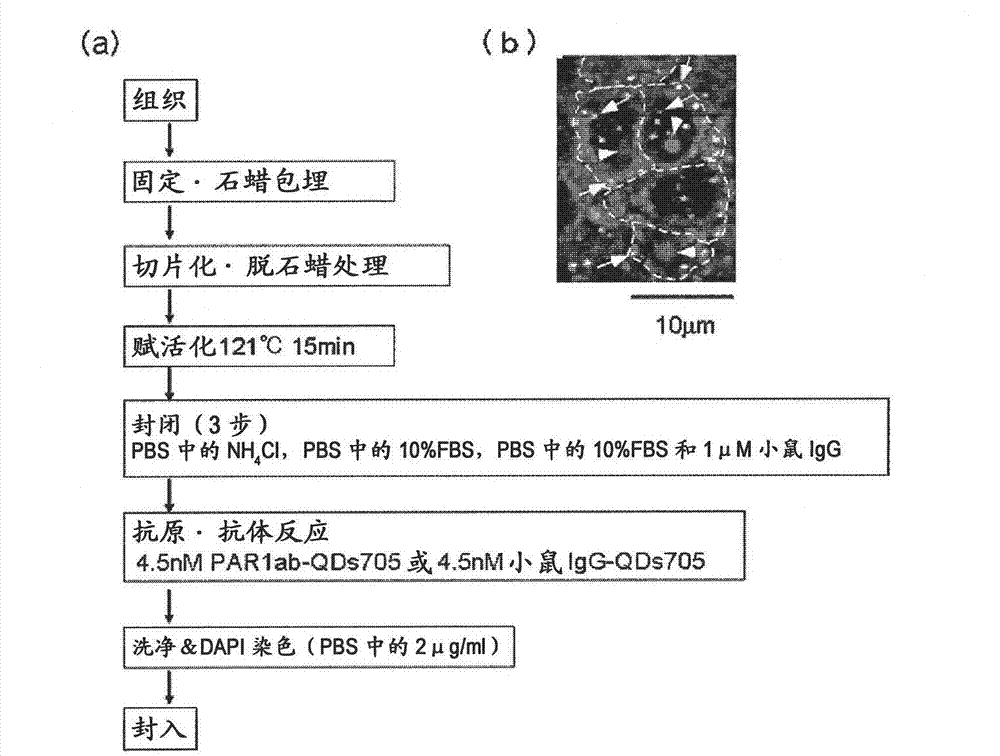
[0087] Preparation method of immunostained tissue sample

[0088] In order to distinguish the difference from Hercep Test (detection of breast cancer tissue by HER2 antibody), all Hercep Test-negative tissue samples were used as human breast cancer pathological tissue. On the other hand, there are cancer cases unrelated to the three main factors (ER, PgR, HER2) related to the proliferation of breast cancer. It is generally believed that the "triple negative" breast cancer has a poor prognosis and is difficult to treat. Here, "triple negative" breast cancer tissue samples are also discussed. Specifically, 4 test samples of breast cancer tissues from recurrent patients (relapse within about 1 year and death within 4 years) (one of the test samples was triple negative [because 20-25% of recurrent patients are "triple negative", so random Add the "triple negative" label to 1 of the 4 test samples]), "triple negative" breast cancer tissue from recurrent patients (recurrence within...

Embodiment 2

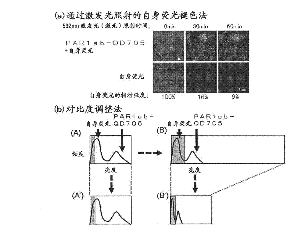
[0090] Problems with fluorescent immunohistostaining

[0091] For the immunostained tissue sample prepared in Example 1 above, a combination of a confocal unit (manufactured by Yokogawa Electric Corporation), a fluorescence microscope (manufactured by Olympus Corporation), and an electron multiplier CCD (EM-CCD) imaging device (Andor (manufactured by the company) was irradiated with excitation light at 488 nm, and then a fluorescence image (fluorescence still image) of quantum dot fluorescent particles (detected by PAR1ab-QD705) was obtained using a bandpass filter of 695 to 740 nm. figure 1 (b) shows an example of a fluorescence still image of a tissue sample of a patient with recurrent breast cancer. Autofluorescence is so strong that quantum dot fluorescent particles (detected by PAR1ab-QD705) cannot be detected at first glance. Actually, when comparing the fluorescence brightness of autofluorescence and quantum dot fluorescent particles (detected by PAR1-antibody), only a...

Embodiment 3

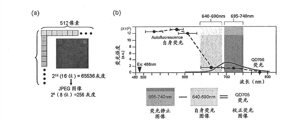
[0094] Creation of corrected fluorescence images with autofluorescence removed

[0095] Fluorescence still images with zero (0) fluorescence intensity of autofluorescence as background are required. In this way, the total fluorescence in the fluorescence still image can be calculated as the fluorescence derived from the quantum dot fluorescent particles. The contrast adjustment method of the above-mentioned embodiment 2 is a division method of fluorescence intensity, and zero (0) cannot be obtained by division, so it must be an image processing method using a subtraction method that can obtain zero (0). Thus, as an image processing method for removing autofluorescence in a fluorescent still image, the method described below is considered. First, the breast cancer tissue sample immunostained with quantum dot fluorescent particles is irradiated with excitation light (laser) with an excitation wavelength of 488 nm, and a fluorescence image of quantum dot fluorescent particles is...


PUM

PropertyMeasurementUnit
wavelengthaaaaaaaaaa

Abstract

Provided is a method for highly accurately and quantitatively determining cancer onset or cancer onset risk by quantitative tissue-staining which is conducted in a biotissue with the use of an antibody capable of recognizing a cancer proliferation inhibitor or a cancer metastasis inhibitor, for example, PAR1 antibody inhibiting the mobility and infiltrating properties of cancer cells. Cancer onset or cancer onset risk is determined by a tissue staining method, said method comprising: a step for labeling an antibody, which is capable of recognizing a cancer proliferation inhibitor or a cancer metastasis inhibitor, with a fluorescent substance and then contacting the fluorescent-labeled antibody with a tissue sample; a step for irradiating a tissue site, which has been contacted with said antibody, with an excitation light to give a fluorescence image; a step for acquiring an intrinsic fluorescence image in a neighborhood region on a shorter or longer wavelength side, compared with the region wherein the wavelength of the fluorescence emitted by the fluorescent substance is acquired, in the same visual field at the same focus point as said fluorescence image; a step for conducting an image processing for removing the fluorescent brightness of said intrinsic fluorescence image from the fluorescent brightness of said fluorescence image to give a corrected fluorescence image; a step for counting cells at the tissue site which has been contacted with the antibody; a step for measuring the average fluorescent brightness per fluorescent particle; and a step for calculating the fluorescent particle count per cell.

Description

technical field [0001] The present invention relates to a method for determining the onset of cancer or the risk of developing cancer, using a tissue staining method that effectively eliminates the influence of autofluorescence and enables quantitative analysis with high precision. Background technique [0002] Cancer, together with vascular system diseases represented by myocardial infarction and cerebral infarction, are the two major causes of death in adults. For example, although the incidence rate of breast cancer in Japan is lower than that of European and American countries, it tends to increase in recent years. In 1998, it surpassed the incidence rate of gastric cancer and became the first female incidence rate. According to the statistics of the Ministry of Health, Labor and Welfare of Japan in 2005, which is the most recent report, the annual number of breast cancer patients exceeds 50,000. [0003] In the diagnosis of cancer, in addition to image diagnosis such a...

Claims


Application Information

Patent Timeline
Patent Type & Authority Applications(China)
IPC IPC(8): G01N33/574G01N21/64G01N33/48G01N33/53G01N33/536
CPCG01N21/6428G01N33/57415G01N2800/50G01N21/6458G01N33/588B82Y15/00G01N21/6486
Inventor 权田幸祐宫下穰武田元博大内宪明
Owner TOHOKU UNIV
Why Eureka
Try Eureka