Method and composition for optical clearing of tissues

a tissue and optical clearing technology, applied in the field of tissue optical clearing, to achieve the effect of excellent tissue transparency, high resolution, and convenient us

Active Publication Date: 2020-07-02
UNITED STATES OF AMERICA
View PDF0 Cites 1 Cited by
  • Summary
  • Abstract
  • Description
  • Claims
  • Application Information

AI Technical Summary

Benefits of technology

[0010]Organ homeostasis, cellular differentiation, signal relay, and in situ function all depend on the spatial organization of cells in complex tissues. For this reason, comprehensive, high resolution mapping of cell positioning, phenotypic identity, and biological state in the context of macro-scale tissue structure is critical to a deeper understanding of developmental processes as well as physiological and patho-physiological processes. The present invention is based on a surprising

Problems solved by technology

The discovery is unexpected, in part, because many other compounds did not provide adequate clearing results a

Method used


Image

  • Method and composition for optical clearing of tissues
  • Method and composition for optical clearing of tissues
  • Method and composition for optical clearing of tissues

Examples


example 1

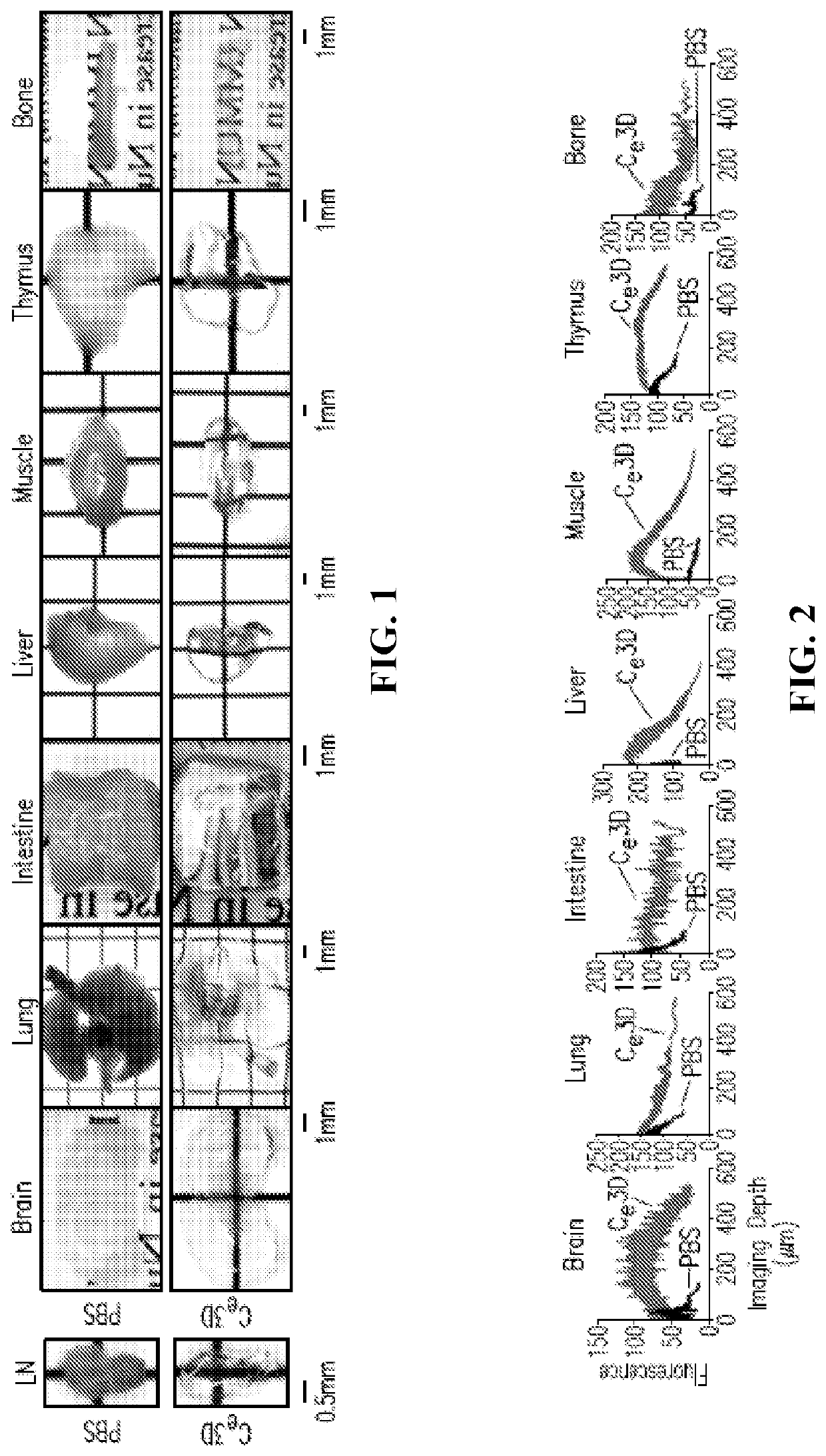
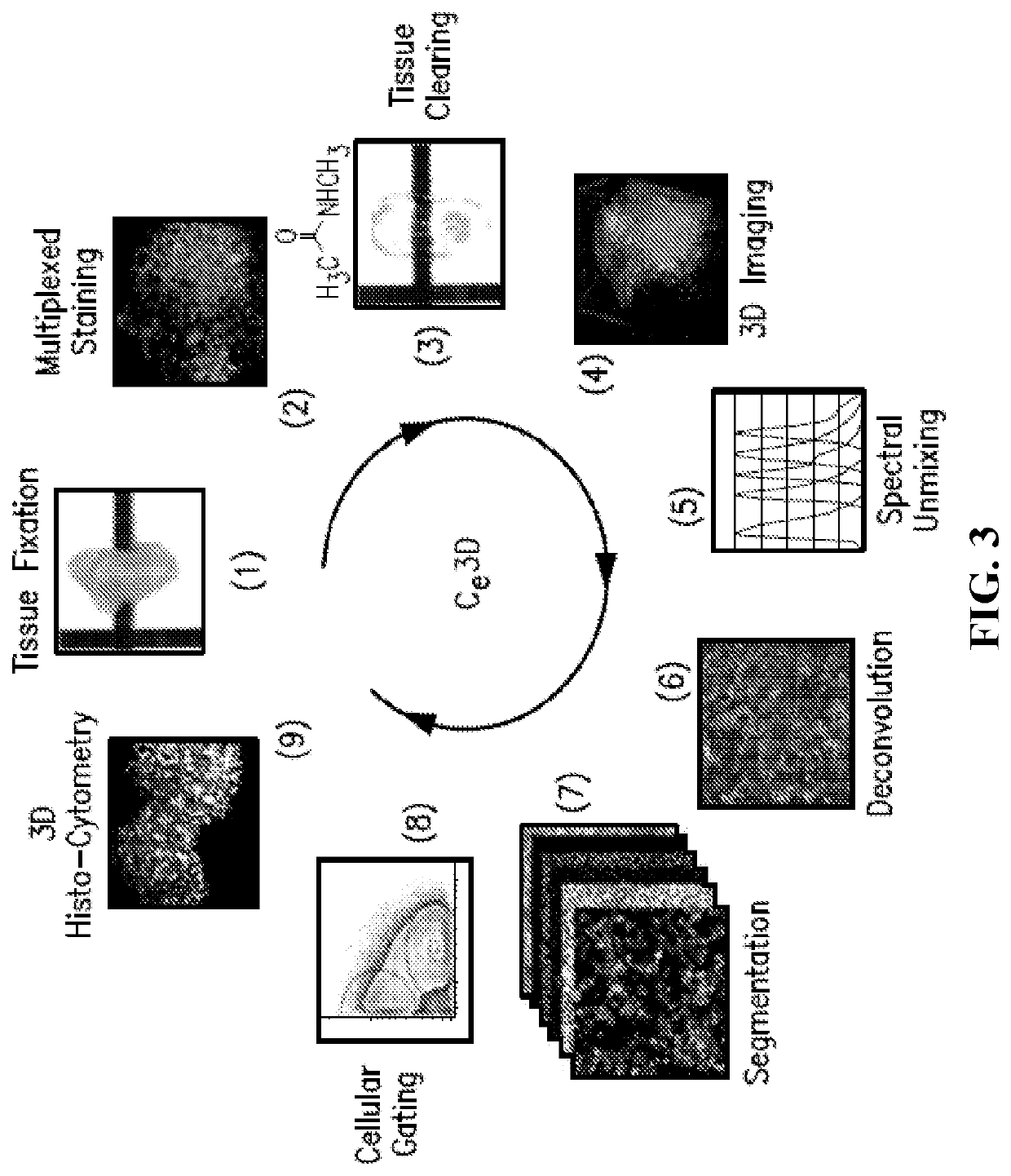
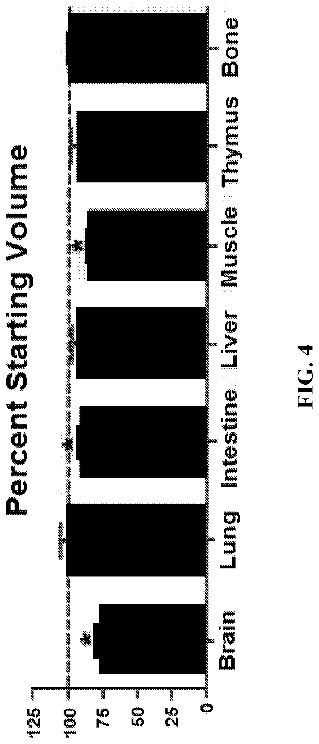
[0161]This example demonstrates the improved clearing of tissues, including preservation of cellular morphology, reporter fluorescence and epitope labeling, when tissues are treated in accordance with embodiments of the invention. In this example, mouse tissues were cleared, stained, and imaged in accordance with embodiments of the invention. The specific details of this experiment are as follows.

[0162]Mice: CD11c-YFP (B6.Cg-Tg(Itgax-Venus)1Mnz / J), actin-DsRed (B6.Cg-Tg(CAG-DsRed*MST)1Nagy), Cxcl12-DsRed (Cxcl12tm2.1Sjm / J), Cx3cr1-GFP (B6.129P-Cx3cr1 tm1Litt / J) and Histone GFP-tagged reporters (B6.Cg-Tg(HIST1H2BB / EGFP)1Pa) were obtained from Jackson Laboratories. Fluorescent Confetti animals were generated by crossing B6.129P2-Gt(ROSA)26Sortm1(CAG-Brainbow2.1)Cle / J×B6.Cg-Tg(UBC-cre / ERT2)1Ejb / 2J Jackson Laboratories). Mice heterozygous for both transgenes were injected intraperitoneally with tamoxifen 100 micrograms per gram of body weight in peanut oil (Sigma Aldrich) for five conse...

example 2

[0175]This example demonstrates an improved method of clearing of lipid-rich tissues, including preservation of cellular morphology, reporter fluorescence and epitope labeling, when tissues are treated in accordance with an embodiment of the invention.

[0176]In this example, mouse brain tissue was cleared, stained, and imaged in accordance with Example 1 except as indicated for the following: (1) 1×BD Perm / Wash™ buffer (available from BD Biosciences) was used instead of BD Peimeabilization buffer for tissue washing; and (2) 1% BSA and normal mouse serum in 1×BD Perm / Wash™ was used as the blocking and antibody dilution buffer. BD Penn / Wash™ is a saponin containing buffer which also includes sodium azide and Fetal Bovine Serum.

[0177]This method provides improved clearing of tissue that are lipid-rich, such as brain tissue, by using a commercially available (e.g., one available from BD Biosciences) saponin-based surfactant.

example 3

[0178]This example demonstrates the improved clearing of fat-rich tissues, including preservation of cellular morphology, reporter fluorescence and epitope labeling, when tissues are treated in accordance with an embodiment of the invention.

[0179]In this example, mouse mammary gland tissue was cleared, stained, and imaged in accordance with Example 1 except as indicated below.

[0180]After fixation, samples were incubated at room temperature for one hour each in 50%, 80%, 95%, 100% of acetone (acetone was diluted in water). The incubation 100% acetone can be extended to 1 to 3 days (at room temperature) depending on the size of sample. Then the samples were incubated in 95%, 80%, 50% of acetone for 1 hour at room temperature, and finally in PBS.

[0181]The 3D structure of ducts of the mouse mammary glands were visualized by Ce3 D clearing. The directly conjugated antibody (Alexa Fluor 647) for E-cadherin was used to stain ducts, dendritic cells were visualized by endogenous expression o...


PUM


Abstract

Disclosed are compositions, methods, and kits for clearing tissue that preserve cellular morphology, reporter fluorescence, and epitope labeling which allow for quantitative phenotypic analysis of intact organs. The compositions include, for example, a compound of formula R1—C(X)—NR2R3, wherein R1 is alkyl, haloalkyl, hydroxyalkyl, amino, or alkylamino, X is O or S, and R2 and R3 are independently H, alkyl, or hydroxyalkyl, a salt thereof, or a combination thereof, and at least one non-ionic density gradient medium. Also disclosed are methods for clearing tissue comprising positioning a tissue in a tissue clearing composition and allowing a tissue clearing composition to permeate the tissue. Further disclosed are methods for visualizing tissue characteristics which involve fixing a tissue, staining the tissue, positioning the tissue in the tissue clearing composition and allowing the tissue clearing composition to permeate the tissue, and imaging the tissue utilizing a microscope or tissue scanning device.

Description

CROSS-REFERENCE TO A RELATED APPLICATION[0001]This patent application claims the benefit of U.S. Provisional Patent Application No. 62 / 380,593 filed Aug. 29, 2016, which is incorporated herein by reference in its entirety for all purposes.BACKGROUND OF THE INVENTION[0002]Major physiological processes rely on the precise positioning of diverse cell types in specific anatomical locations. Such organization allows for exposure of cells to appropriate tissue microenvironments that shape their differentiation, promote appropriate cell-cell communication events, and collectively define the global properties of the whole organ. Understanding these structure-function relationships requires a detailed mapping of both the large-scale organization and fine-grained molecular and cellular composition of complex tissues.[0003]The majority of information to date of such organization at the cell and subcellular levels comes from microscopic examination of relatively thin (e.g., 5 to 20 micrometer) ...

Claims


Application Information

Patent Timeline
IPC IPC(8): G01N1/30
CPCG01N2001/302G01N1/30
Inventor GERMAIN, RONALD N.LI, WEIZHEGERNER, MICHAEL Y.
Owner UNITED STATES OF AMERICA
Why Eureka
Try Eureka