
Method for removing ring artifact of CT (computed tomography) reconstructed image

A technology for ring artifact and image reconstruction, applied in the field of medical image processing, can solve the problems of affecting image quality, long time to remove ring artifact, less number of ring artifact, etc., so as to improve quality and reduce ring artifact. The effect of reducing the shadow area and reducing the removal time

Active Publication Date: 2013-07-03
SHANGHAI UNITED IMAGING HEALTHCARE
View PDF2 Cites 6 Cited by
  • Summary
  • Abstract
  • Description
  • Claims
  • Application Information

AI Technical Summary

Problems solved by technology

In the existing method of removing ring artifacts, all images are processed to obtain ring artifact information of all images, but in the process of post-processing, the number of ring artifacts is often relatively small, and only in the image Some few parts appear, and the rest are free of ring artifacts, which often results in a longer time to remove ring artifacts
In addition, in the existing method, it is sometimes possible to detect the error value of the ring artifact in the area without the ring artifact in the image, so that the error that should not be removed is mistakenly removed from the original image, which affects the image quality. quality

Method used


Image

  • Method for removing ring artifact of CT (computed tomography) reconstructed image
  • Method for removing ring artifact of CT (computed tomography) reconstructed image
  • Method for removing ring artifact of CT (computed tomography) reconstructed image

Examples


Embodiment Construction

[0023] The present invention will be further described below in conjunction with the accompanying drawings and embodiments.

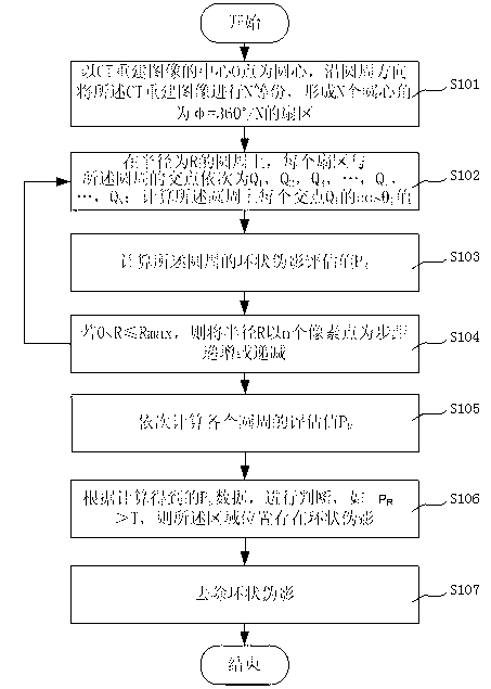
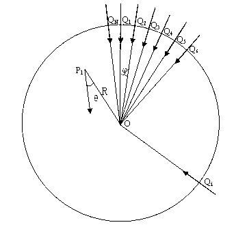
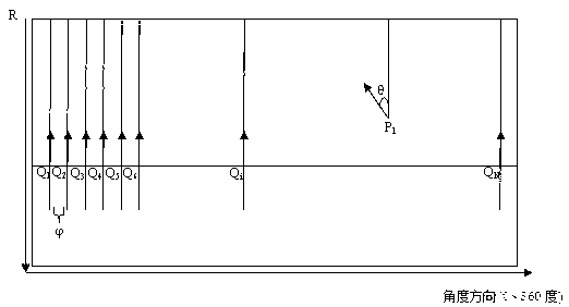
[0024] figure 1 A flow chart of removing ring artifacts in CT reconstruction images for the present invention; figure 2 Schematic diagram for positioning ring artifacts, image 3 for right figure 2 Schematic diagram of coordinates after polar coordinate conversion.

[0025] Please refer to figure 1 , figure 2 with image 3 , the removal method of the CT reconstruction image ring artifact provided by the present invention, comprises the steps:

[0026] Step S101, taking the center O of the CT reconstruction image as the center of the circle, dividing the CT reconstruction image into N equal parts along the circumferential direction to form N sectors with a central angle of φ=360° / N, wherein N is a positive integer, Preferably, N is a positive integer greater than or equal to 90, that is, the central angle Take 0 to 4 degrees.

[0027] Step S...


PUM


Abstract

The invention discloses a method for removing a ring artifact of a CT (computed tomography) reconstructed image. The method comprises the following steps of: a, taking a center O point of the CT reconstructed image as a circle center; dividing the CT reconstructed image into N equal parts along the peripheral direction; b, setting the intersection points of all sector and the circumference orderly as Q1, Q2, Q3,..., and QN on a circumference of which the radius is R, calculating the value of each intersection point Qi on the circumference; c, calculating a ring artifact evaluation value PR of the circumference; d, if R is smaller than or equal to Rmax and greater than 0, gradually increasing or gradually reducing the radius R by taking n pixel points as step pitches; e, repeating the steps b, c and d, orderly calculating the evaluation value PR of each circumference; and f, judging according to calculated PR data, if an absolute value of PR is greater than T, judging that no ring artifact exists in an area, and g, removing the ring artifact. By adopting the removing method provided by the invention, the time spent on removing the ring artifact can be reduced; and the false detection of a ring artifact area can be reduced to a certain extent.

Description

technical field [0001] The invention relates to medical image processing technology, in particular to a method for removing ring artifacts in CT reconstructed images. Background technique [0002] Since the advent of CT in the 1970s, it has been continuously improved. From the first generation to the fifth generation, the scanning time has been continuously shortened and the image quality has been improved. CT has been widely used in many fields of medical diagnosis. CT measures the cumulative attenuation coefficient (or projection) of X-rays passing through the human body in all directions, and then calculates the distribution of X-ray attenuation coefficients on the entire section by computer, and finally displays them in the form of images to assist clinical Make a diagnosis of the disease. Because it can provide higher soft tissue resolution than ordinary X-ray imaging and solve the problem of three-dimensional structure overlap, it has epoch-making significance in med...

Claims


Application Information

Patent Timeline
Patent Type & Authority Applications(China)
IPC IPC(8): G06T5/00
Inventor 不公告发明人
Owner SHANGHAI UNITED IMAGING HEALTHCARE
Learn More