
Three-dimensional model to two-dimensional image space registering method of human body bone joint system

A three-dimensional model, spatial registration technology, applied in image analysis, image data processing, instruments, etc., can solve the problems of registration accuracy limitation, time-consuming, cumbersome operation, etc., to increase accuracy and reliability, The effect of reducing time cost and improving work efficiency

Inactive Publication Date: 2013-08-14
THE FIRST AFFILIATED HOSPITAL OF THIRD MILITARY MEDICAL UNIVERSITY OF PLA
View PDF2 Cites 13 Cited by
  • Summary
  • Abstract
  • Description
  • Claims
  • Application Information

AI Technical Summary

Problems solved by technology

The whole process is cumbersome and time-consuming
Since the comparison of the contour lines is completely recognized by the naked eye, the matching result depends on the subjective judgment of the operator and lacks quantitative indicators, so the accuracy of the registration is limited, and it is easily affected by the operator's technology, with large individual differences.
These shortcomings limit its application in bone and joint kinematics detection
At present, the two-plane X-ray image monitoring and model registration method can only be carried out on a small scale in the laboratory, and cannot be widely used in clinical practice.

Method used


Image

  • Three-dimensional model to two-dimensional image space registering method of human body bone joint system
  • Three-dimensional model to two-dimensional image space registering method of human body bone joint system
  • Three-dimensional model to two-dimensional image space registering method of human body bone joint system

Examples


Embodiment 1

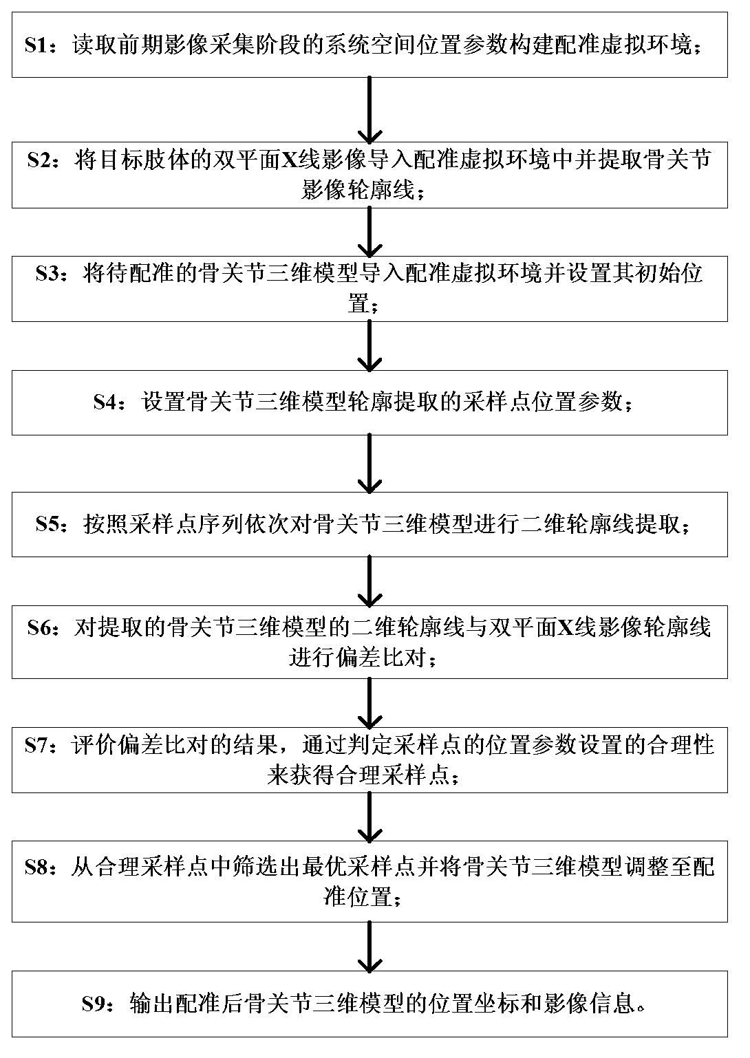
[0041] figure 1 It is a flow chart of Embodiment 1 of the registration method, as shown in the figure, the three-dimensional model of the human bone joint system-two-dimensional image space registration method provided by the present invention includes the following steps:

[0042] S1: Read the system spatial position parameters in the previous image acquisition stage to construct a registration virtual environment; the registration virtual environment is the same as the real physical environment of biplane X-ray image acquisition, and is established through the following steps:

[0043] S11: Read the position information of the X-ray emitting device and the X-ray receiving device in the dual-plane X-ray image acquisition device, and construct a spatial coordinate system;

[0044] S12: Configure the initial placement point of the virtual camera in the space coordinate system according to the position information of the X-ray emitting device, and configure the initial placement...

Embodiment 2

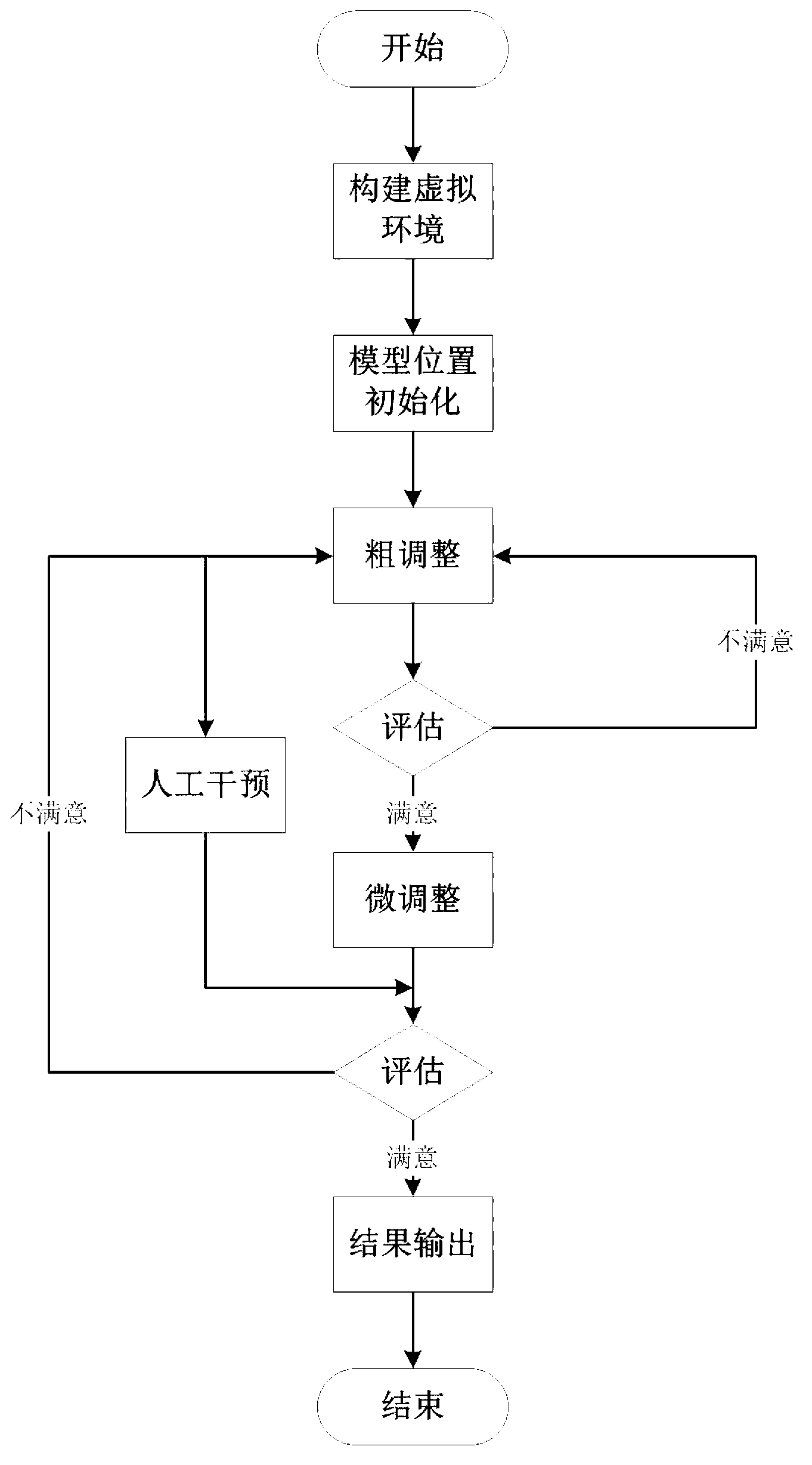
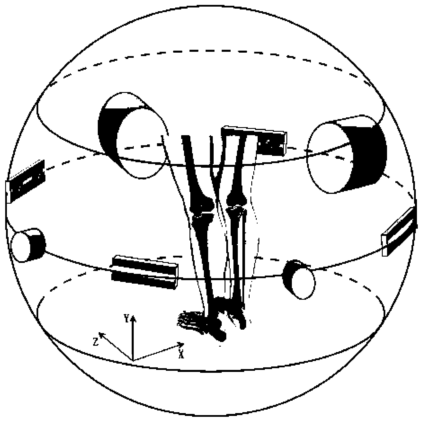
[0056] figure 2 It is the flow chart of Embodiment 2 of the registration method, image 3 It is a schematic diagram of the integrated acquisition of dual-plane X-ray images and somatosensory motion in the early stage. Figure 4 Schematic diagram of the initial alignment between the virtual environment construction and the bone joint model for registration, Figure 5 Schematic diagram for X-ray image contour extraction, Figure 6 Extract schematics for model contour projection, Figure 7 extract the schematic for the model outline, Figure 8 Schematic extraction for coarse-tuned sampling of model contours, Figure 8 The black dots in represent the distribution state of coarse adjustment sampling points, and the three-dimensional model of the bone joint system is placed in the middle. Figure 9 Schematic extraction for fine-tuning sampling of model contours, Figure 9 The black dots in the figure indicate the distribution state of the fine-tuning sampling points, and the ...


PUM


Abstract

The invention discloses a three-dimensional model to two-dimensional image space registering method of a human body bone joint system. Firstly, system position parameters at a bi-plane X-ray image collecting phase are obtained to construct a registering virtual environment; secondly, limb somatosensory motion information is utilized to initialize and align a bone joint three-dimensional model in the space; thirdly, space sampling points are set, and two-dimensional contour line sampling and extraction are carried out in sequence on the three-dimensional model; fourthly, deviation comparison is carried out on three-dimensional model contour lines and X-ray image contour lines, and optimal contour lines and corresponding sampling points are screened; and fifthly, the bone joint three-dimensional model is adjusted until registering is finished through the spatial position information of the optimal sampling points. According to the method, the rapid registering between the three-dimensional model and two-dimensional images of a bone joint system is realized under the condition of less manual intervention, complicated manual manipulation can be replaced, the efficiency and the registering precision are improved, and labor cost and time cost are saved.

Description

technical field [0001] The invention relates to the field of medical equipment, in particular to a method for registering a three-dimensional model of a human bone joint system and a two-dimensional image. Background technique [0002] The occurrence and development of bone and joint injuries and diseases are often accompanied by changes in local joint motion characteristics. At the same time, some abnormal joint activities can also lead to diseases. Detecting the kinematic characteristics of the bone and joint system in vivo is of great significance to the scientific research, diagnosis, treatment and prognosis evaluation of diseases. [0003] Optical motion capture and motion sensing are currently widely used kinematic detection methods. However, in these methods, optical markers or physical sensors that capture motion are installed on the surface of the limbs. Due to the large relative movement between the skin and bony structures, the limb movement information obtained...

Claims


Application Information

Patent Timeline
Patent Type & Authority Applications(China)
IPC IPC(8): G06T7/00
Inventor 尹力杨柳
Owner THE FIRST AFFILIATED HOSPITAL OF THIRD MILITARY MEDICAL UNIVERSITY OF PLA
Learn More