

Application of FITC-dextran in manufacturing fundus angiographic agent
A technology of angiography and dextran, which is applied in the field of application of FITC-dextran in the preparation of fundus angiography, can solve the problems of application limitations of ICG fundus angiography, and achieve high resolution and high definition Effect
- Summary
- Abstract
- Description
- Claims
- Application Information
AI Technical Summary
Problems solved by technology
Method used
Image

Examples
Embodiment 1
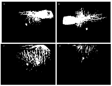
[0064] FITC-dextran fundus angiography: Simultaneous observation of retinal and choroidal vessels in the same fundus angiogram image.
[0065] Materials and methods: The test subjects were healthy rabbits weighing 1.8-2.5 kg. FITC-dextran (FD-70S, Sigma, St. Louis, MO) with an average molecular weight of 70 kDa was used as a contrast agent. Dissolve FITC-dextran in water to obtain a solution with a concentration of 25 mg / ml. Each rabbit was intravenously injected with 0.3 ml of FITC-dextran solution. Angiographic images were acquired using a conventional fundus fluorescence system (Topcon 50 VT).
[0066] figure 1 The results in show FICT-dextran fundus angiography images of a normal rabbit eye. figure 1 Red arrows in A, B, and C indicate normal retinal vessels, in figure 1 Yellow arrows in A, B, C, and D indicate normal choroidal vessels, suggesting that FITC-dextran angiography allows simultaneous visualization of retinal and choroidal vessels in the same fundus angi...
Embodiment 2
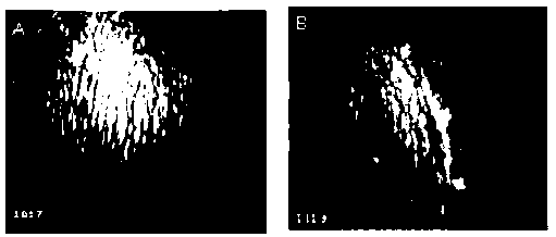
[0068] Effective Imaging Duration of Rabbit FITC-dextran Fundus Angiography
[0069]Materials and methods: FITC-dextran with an average molecular weight of 70 kDa (FD-70S, Sigma Corporation, St. Louis, Missouri) was dissolved in water for injection to a final concentration of 25 mg / ml. Dutch Belted rabbits were injected with a volume of 0.3 ml of the above solution through the marginal vein. Fluorescence images of choroidal vessels were monitored for at least 2 hours after injection of FITC-dextran solution, and choroidal angiography images were acquired using a conventional fundus fluorescence imaging system (Topcon 50 VT).
[0070] figure 2 Fluorescence images of choroidal vessels within 2 hours after a single intravenous injection of FITC-dextran solution are shown. figure 2 Panels A to H show contrast images of normal choroidal vessels acquired at 10 s, 30 s, 1 min, 1.4 min, 10.4 min, 32.6 min, 59.3 min, and 119.4 min after intravenous injection of FITC-dextran.
[00...
Embodiment 3
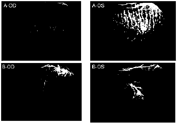
[0073] Application of FITC-dextran fundus angiography in rabbit eyes with retinal detachment
[0074] Materials and methods: Retinal detachment was induced by subretinal injection of Healon in the eyes of pigmented rabbits. FITC-dextran (FD-70S, Sigma, St. Louis, MO) with an average molecular weight of 70 kDa was dissolved in water for injection to a final concentration of 25 mg / ml. Rabbits were injected intravenously with a volume of 0.3 ml of the above solution through the marginal vein. Choroidal angiography images were acquired using a conventional fundus fluorescence system (Topcon 50VT).
[0075] Fundus angiography images of retinal detachment in rabbit eyes were obtained by FITC-dextran fundus angiography. image 3 The yellow arrow in indicates the area of retinal detachment with sharp edges, and the image also shows fluid in the subretinal space at the site of retinal detachment and choroidal vessels underlying the area of retinal detachment.
[0076] FITC-dextr...
PUM
 Abstract
Description
Claims
Application Information