Photoactivated single-molecular fluorescence microscope for biochemical reaction kinetics and test method
A biochemical reaction and single-molecule technology, applied in the field of biochemical experiments, can solve the problems of technical implementation difficulties and lack of means
- Summary
- Abstract
- Description
- Claims
- Application Information
AI Technical Summary
Problems solved by technology
Method used
Image
Examples
Embodiment 1
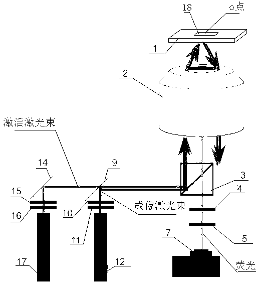
[0022] This embodiment provides a light-activated single-molecule fluorescence biochemical reaction dynamics microscope, which includes a sample stage 1 , a microscope objective lens 2 , an imaging laser emitter 12 , a camera 7 and an activation laser emitter 17 . The upper surface of the sample stage 1 carries a light-transmitting substrate 18 , the sample stage 1 has a through hole passing through the upper and lower surfaces, and the light-transmitting substrate 18 covers the through hole. In this embodiment, the transparent substrate 18 is a glass slide with flat upper and lower surfaces.
[0023] The upper surface of the light-transmitting substrate 18 carries material molecules A and material molecules B that biochemically react when in contact with each other, wherein the material molecules A are fixed on the upper surface of the light-transmitting substrate 18, and the material molecules Molecule B itself is an activatable fluorescent molecule or labeled with an activa...
Embodiment 2
[0032] The method of using the light-activated single-molecule fluorescent biochemical reaction kinetic microscope described in Example 1 to conduct experiments and observe the dynamic process of the interaction between E-Cadherin molecules includes the following steps:
[0033] 1) Immobilizing the substance A, E-Cadherin molecule, participating in the target reaction on the upper surface of the light-transmitting substrate 18 . In this embodiment, the carbon terminal of E-Cadherin will be passed through (HIS) 6 group connected to the NTA-Ni on the upper surface of the light-transmitting substrate 18 2+ on the spot. In the experiment, the distribution density of E-Cadherin molecules on the surface is about 2 per square micron.
[0034] 2) Substance B participating in the target reaction, molecules composed of E-Cadherin and GFP, is added dropwise to the solution on the light-transmitting substrate 18 immobilized with E-Cadherin molecules. In this embodiment, the solution ...
Embodiment 3
[0038] The method of using a light-activated single-molecule fluorescent biochemical reaction kinetic microscope described in Example 1 to conduct experiments to observe the dynamic process of the interaction between Calmodulin (Calmodulin) and the target polypeptide molecule includes the following steps:
[0039] 1) Immobilizing the substance A involved in the target reaction, calmodulin molecule, on the upper surface of the light-transmitting substrate 18 . In this embodiment, the nitrogen terminal of the calmodulin molecule will be passed through (HIS) 6 group connected to the NTA-Ni on the upper surface of the light-transmitting substrate 18 2+ on the spot. In the experiment, the distribution density of calmodulin molecules on the surface was about 2 per square micron.
[0040] 2) Substance B participating in the target reaction, C28W molecules labeled with Alex488, is added dropwise to the solution on the light-transmitting substrate 18 immobilized with calmodulin mol...
PUM
| Property | Measurement | Unit |
|---|---|---|
| thickness | aaaaa | aaaaa |
Abstract
Description
Claims
Application Information
 - R&D
- Intellectual Property
- Life Sciences
- Materials
- Tech Scout
- Unparalleled Data Quality
- Higher Quality Content
- 60% Fewer Hallucinations
Browse by: Latest US Patents, China's latest patents, Technical Efficacy Thesaurus, Application Domain, Technology Topic, Popular Technical Reports.
© 2025 PatSnap. All rights reserved.Legal|Privacy policy|Modern Slavery Act Transparency Statement|Sitemap|About US| Contact US: help@patsnap.com

