Method for differential counting of white blood cells based on morphology

A technology based on morphology, classification and counting, applied in the field of image processing, can solve problems such as subjective factors, abnormal judgment or identification that cannot be diversified, and large detection workload, so as to improve the speed and accuracy of diagnosis, and achieve rapid Accurate classification and the effect of overcoming subjective factors

Inactive Publication Date: 2013-10-09
SUZHOU CHUANGJI BIOLOGICAL TECH
View PDF2 Cites 22 Cited by
  • Summary
  • Abstract
  • Description
  • Claims
  • Application Information

AI Technical Summary

Problems solved by technology

[0003] Manual classification and counting of white blood cells in peripheral blood smears after Wright staining is a routine work in clinical testing at present. Due to the heavy workload, long testing time, cumbersome process, and low work efficiency, the analysis of cells is subject to testing. Limitations of physician experience and visual resolution, too many subjective factors, lack of objective standards
[0004] Although the automatic blood cell analyzer can quickly perform white blood cell classification and counting, it has the following shortcomings that are difficult to overcome: ① Cells have to pass through very small holes, which is easy to cause blockage; ② There is no ability to identify abnormal cells, which is not conducive to clinical diagnosis; ③ Cannot preserve the original physiological state of the sample
However, pattern recognition has its limitations in medical applications. Medical detection mainly detects abnormal states (abnormal conditions that occur in the case of disease), and these abnormal states are extremely diverse, and pattern recognition is Based on a non-measurement comparison algorithm compared with normal samples, it cannot be used to judge or identify abnormalities in diversity

Method used


Image

  • Method for differential counting of white blood cells based on morphology
  • Method for differential counting of white blood cells based on morphology
  • Method for differential counting of white blood cells based on morphology

Examples


Embodiment

[0035] (1) After the peripheral blood smear was stained by Wright's, the image was collected by microscope;

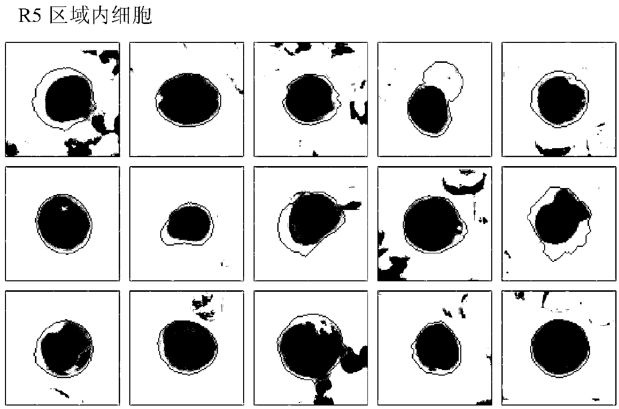
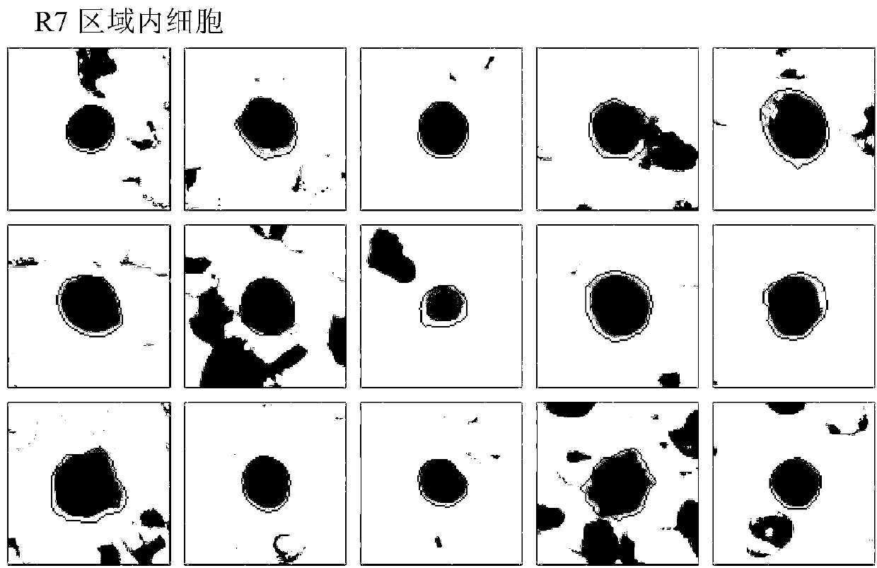
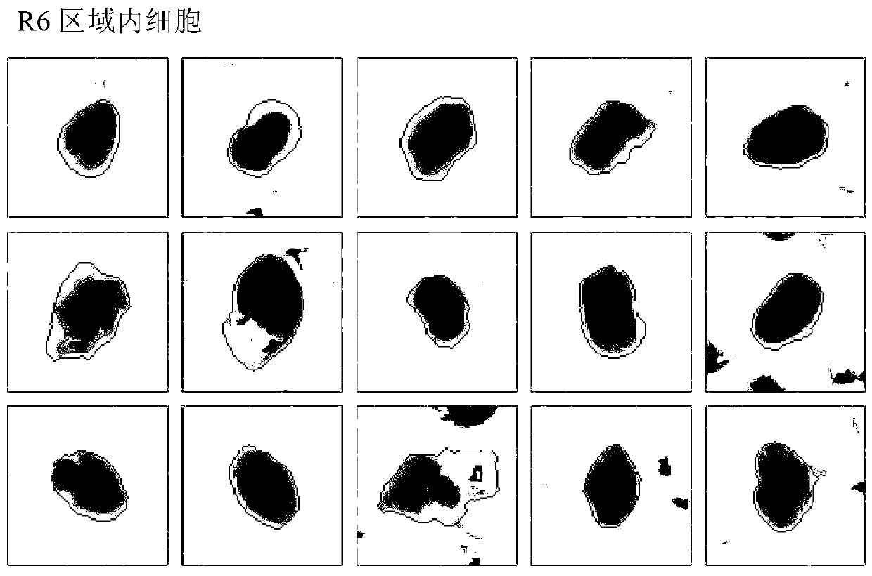
[0036] (2) Input the image into the computer system for processing, see figure 1 , to analyze the morphological parameters of white blood cells, and construct a two-dimensional scatter diagram with the cell nuclear plasma ratio as the abscissa, and construct a two-dimensional scatter diagram with the roundness of the nucleus (nuclei radius variation coefficient) as the ordinate, and construct a two-dimensional scatter diagram according to the corresponding area of ​​the cell Microscopic image, the leukocytes are divided into four areas: lymphocyte area R1, monocyte area R2, granulocyte area R3 and extinct cell area R4;

[0037] (3) Further analyzing the morphological parameters of the regional cell lymphocyte area R1, monocyte area R2, and granulocyte area R3 divided in step (2);

[0038] see figure 2 and 3 , with the total brightness of the nucleus as the abscissa...


PUM


Abstract

The invention discloses a method for differential counting of white blood cells based on morphology. The method includes the steps that after Wright stain is conducted on peripheral blood smears, a microscope is used for collecting an image; after the image is processed by a computer system, morphology parameter analysis is conducted on the white blood cells, and the white blood cells are divided into a lymphocyte area, a monocyte area, a granulocyte area and a cell extinction area; the morphology parameter analysis is conducted on the area cells which are divided in the previous step, and the cells are further divided into monocytes, lymphocytes, eosinophilic granulocytes and neutrophile granulocytes precisely; the differential counting statistics are carried out on the variety of the divided cells. The computer system is used for analyzing the processed image, several simple morphology parameters are used for making a two-dimensional scatter diagram, all the cells are presented in a statistical diagram mode, the white blood cells can be classified rapidly and precisely, white blood cell counting is automatic, compared with manual counting, the procedures are simple and fast, the diagnose speed is greatly increased, and accuracy is improved substantially.

Description

technical field [0001] The invention belongs to the field of image processing, and in particular relates to a white blood cell classification and counting method. Background technique [0002] In a blood sample, the number of white blood cells is only 1 / 800 of that of red blood cells. In a cell image collection field of view, there will be about 1-5 white blood cells. White blood cells are an important part of the body's defense system. Different granules are divided into granulocytes, monocytes and lymphocytes, and granulocytes are divided into eosinophils, basophils and neutrophils according to the nature of the granules. The composition percentage of various types of white blood cells and the total number of white blood cells is an important item in the blood test. After Wright staining, the types of white blood cells can be distinguished according to the nuclear staining, cytoplasmic staining, and nuclear shape of various mature white blood cells under the microscope. ...

Claims


Application Information

Patent Timeline
Patent Type & Authority Applications(China)
IPC IPC(8): G06M11/00G06K9/62G01N15/10
CPCG01N33/56972G01N15/1475
Inventor 殷跃锋
Owner SUZHOU CHUANGJI BIOLOGICAL TECH
Why Eureka
Try Eureka