
Integrated X-ray protection device on angiography machine

An angiography and protective device technology, applied in the field of medical devices, can solve the problems of high loading and unloading costs, increase the risk of surgery, prolong the operation time, etc., and achieve the effect of reducing the space occupancy rate, increasing the risk of the operation, and prolonging the operation time.

Active Publication Date: 2015-01-21
BEIJING ORIENTAL E T MEDICAL EQUIP
View PDF7 Cites 3 Cited by
  • Summary
  • Abstract
  • Description
  • Claims
  • Application Information

AI Technical Summary

Problems solved by technology

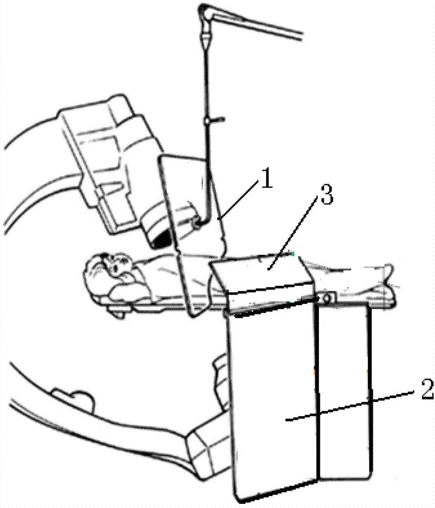
When the bed surface moves, the protective screen 1 cannot move along with the bed, and in the process of moving the catheter bed, the protective screen 1 cannot continue to maintain the pre-adjusted protective position as close as possible to the patient. Therefore, once the catheter bed is moved, not only the The optimal combined positional relationship between the protective screen 1, the protective curtain 2, and the bedside protective board 3, and keeping the protective screen 1 away from the patient, cannot guarantee the protective effect on the secondary rays reflected from the patient's body
[0008] (3) During the operation, after moving the bed surface of the catheter bed each time, in order to avoid the dislocation of the protective device from continuing to provide effective radiation protection, the position of the protective screen has to be readjusted. The protective effect during the adjustment process cannot be guaranteed, and the adjustment The procedure prolongs the operation time, increases the risk of infection for the patient, and increases the risk of the operation
[0009] (4), the protective curtain 2 and the bedside protective plate 3 are immovably fixed on the track beside the bed, and the surgical display parts of the head, chest, abdomen, and lower limbs need to be removed and then reinstalled when the surgical display parts are different, which is complicated and inconvenient to operate , especially when one operation needs to be performed on two parts, the radiation protection of the two parts cannot be considered at the same time, but can only be realized by dividing it into two operations, which increases the cost, time and risk of the operation
[0010] (5) The protective screen 1 is suspended from the ceiling of the catheterization room, so that the loading and unloading of the protective device needs to destroy the ceiling and other infrastructure, which is costly and difficult to install and unload. In particular, once the position is fixed, it is generally difficult to move the protective device to other beds or In the cath room, in order to meet the timely operation, it is necessary to install more protective devices, which increases the equipment expenditure of the hospital, and is likely to cause the problem that some protective devices are idle for a long time and waste hospital space resources

Method used


Image

  • Integrated X-ray protection device on angiography machine
  • Integrated X-ray protection device on angiography machine
  • Integrated X-ray protection device on angiography machine

Examples


Embodiment Construction

[0046] The specific implementation manners of the present invention will be further described in detail below in conjunction with the accompanying drawings and embodiments. The following examples are used to illustrate the present invention, but are not intended to limit the scope of the present invention.

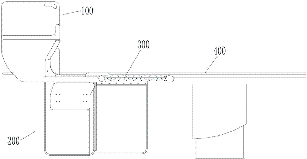
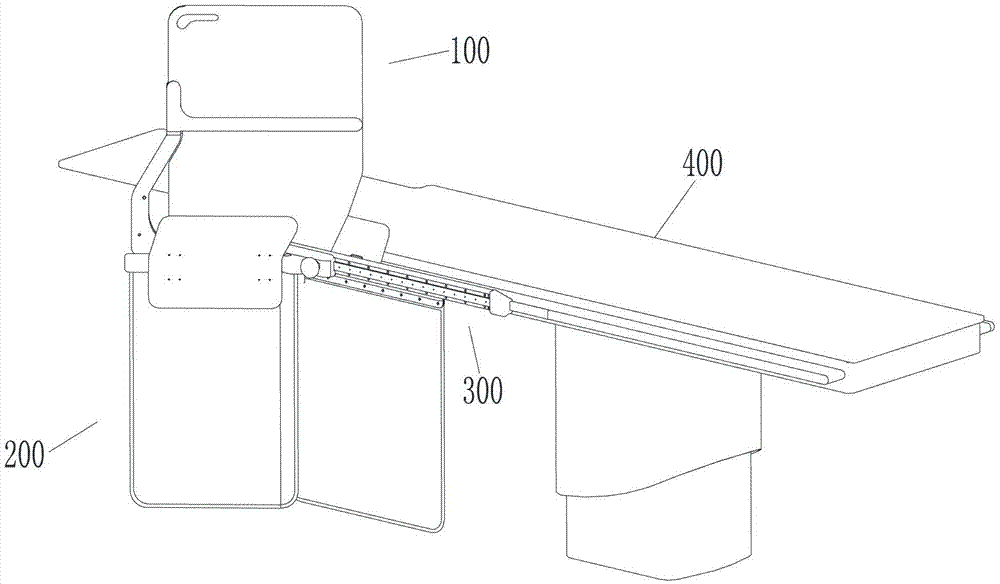
[0047] Such as Figure 2a , 2b , as shown in 2c, an integrated radiation protection device for catheter bed, the device includes a protective screen unit 100 for X-ray protection above the catheter bed 400, a protective screen unit 100 for radiation protection below the catheter bed 400 The curtain unit 200 and the bedside slide rail unit 300 for connecting the side rail 401 of the catheter bed 400, wherein: the protective screen unit 100 is installed on the protective curtain unit 200, for this reason, the protective screen unit 100 and The protective curtain unit 200 is integrated, and this integrated structure can effectively avoid the problem of dislocation of the pr...


PUM


Abstract

The invention relates to the field of medical instruments, in particular to an X-ray protection device with an integrated structure for an angiography machine. The device comprises a protection screen unit, a protection curtain unit and a bedside sliding rail unit, wherein the protection screen unit is arranged on the protection curtain unit, and the protection curtain unit is fixed on the bedside sliding rail unit so as to enable the protection curtain unit and the protection screen unit to synchronously and horizontally move along a bedside. The protection device can synchronously move along with a bed, and in a moving process, the protection device is always in the best protecting state and close to a patient, so that the problem that the radiation, especially the secondary stray radiation caused by the rays reflected by the patient, can not be effectively shielded due to the movement of the bed surface of a conduit bed is effectively solved, and meanwhile, the problems of prolonged surgery time, increased surgery risk and the like that are caused by adjusting the protection screen unit during surgery in the prior art are effectively avoided.

Description

technical field [0001] The invention relates to the field of medical equipment, in particular to an X-ray protection device with an integrated structure used for an angiography machine catheter bed. Background technique [0002] In some clinical operations such as angiography and interventional therapy, it is necessary to rely on X-rays penetrating the patient's body to form images through an angiography machine. In order to avoid leakage of X-ray radiation and protect the safety of doctors, a radiation protection device must be installed next to the angiography machine, and the protection device is generally made of materials with a lead equivalent of 0.5 mm or more to block X-ray radiation. [0003] The clinically required bedside protective device should meet the following requirements: affirmative protective effect, flexible installation, easy operation, does not affect the doctor's operation, and can meet the needs of various positions and positions during the operation...

Claims


Application Information

Patent Timeline
Patent Type & Authority Patents(China)
IPC IPC(8): A61B6/10
CPCA61B6/0407A61B6/107A61B6/4441A61B6/504
Inventor 王盾张汇丰
Owner BEIJING ORIENTAL E T MEDICAL EQUIP
Learn More