Endoscopic device

A technology for endoscopes and image signals, applied in the fields of endoscopes, telescopes, medical science, etc., can solve problems such as blurred blood vessels, complicated operations, and prolonged operation time

Active Publication Date: 2014-01-08
OLYMPUS CORP
View PDF7 Cites 11 Cited by
  • Summary
  • Abstract
  • Description
  • Claims
  • Application Information

AI Technical Summary

Problems solved by technology

In the case of severe bleeding during ESD and other operations, hemostasis must be performed at this time, resulting in prolonged operation time
[0007] However, in order to confirm the position of blood vessels using the above-mentioned infrared endoscope device, as described above, it is necessary to perform complicated work such as intravenous injection of drugs such as ICG.
[0008] In addition, in the case of the above-mentioned infrared endoscope device, the wavelength of the illumination light is the wavelength of near-infrared light, so there is also a problem of blurring blood vessels in the image.

Method used


Image

  • Endoscopic device
  • Endoscopic device
  • Endoscopic device

Examples


no. 1 approach

[0031] (Structure of endoscope device)

[0032] Hereinafter, embodiments of the present invention will be described with reference to the drawings.

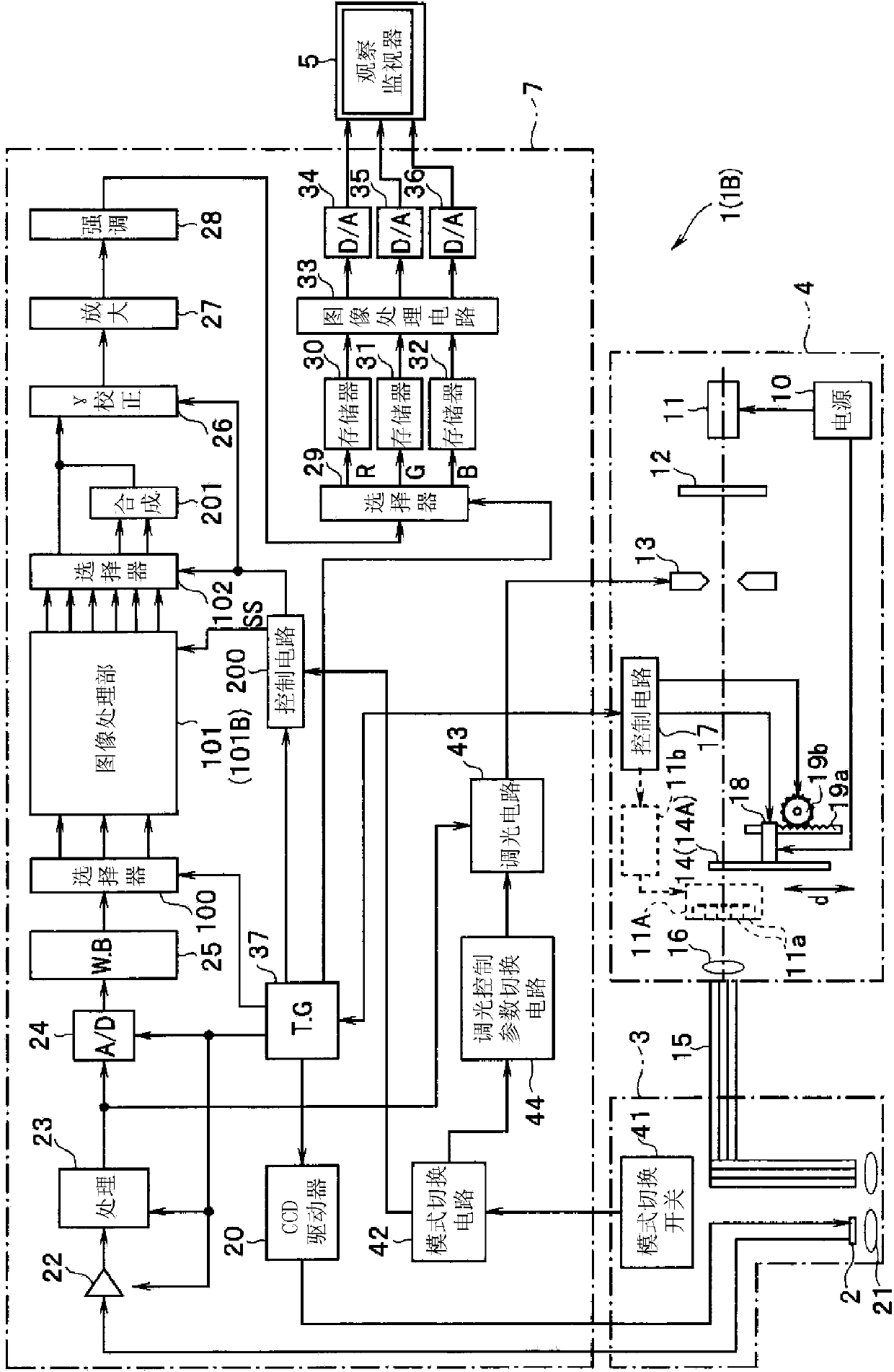
[0033] First, the configuration of the endoscope apparatus according to the present embodiment will be described. figure 1 It is a configuration diagram showing the configuration of the endoscope apparatus according to the present embodiment.

[0034] Such as figure 1 As shown, the endoscope device 1 of this embodiment includes: an electronic endoscope 3, which has a CCD 2 as an imaging element, and the CCD 2 is used as a biological image information acquisition unit inserted into a body cavity to photograph tissues in the body cavity or as a biological a volume image information acquisition unit; a light source device 4, which supplies illumination light to the electronic endoscope 3; and a video processor 7, which performs signal processing on an imaging signal from the CCD 2 of the electronic endoscope 3, and converts the en...

no. 2 approach

[0144] In the first embodiment, at least one narrowband light is used as illumination light to actually irradiate the living body tissue, the image of the returned light is subjected to the above-mentioned frequency band decomposition processing, and at least one frequency band image signal obtained through the frequency band decomposition processing is subjected to emphasis processing, However, in this embodiment, at least one narrow-band light is not actually irradiated to the living tissue, and the image information of the return light of each narrow-band light is obtained by so-called spectral estimation, and the spectral image signal obtained by this spectral estimation is subjected to the above-mentioned frequency band processing. Decomposition processing, performing emphasis processing on at least one frequency band image signal obtained through the frequency band decomposition processing. That is, in the above-mentioned first embodiment, at least one narrow-band light i...

no. 3 approach

[0183] In the first embodiment, at least one narrowband light is actually irradiated to the living body tissue as illumination light, the above-mentioned frequency band decomposition processing is performed on the image of the returned light, and at least one frequency band image signal obtained by the frequency band decomposition processing is subjected to emphasis processing. , in the second embodiment, at least one narrow-band light is not actually irradiated to the biological tissue, but the image information of the return light of each narrow-band light is obtained by so-called spectral estimation, and the spectrum of each wavelength obtained by the spectral estimation is The estimated image signal is subjected to the above-mentioned frequency band decomposition processing and emphasis processing, but in the third embodiment, the image signal of the return light of the actual illumination light of at least two narrow-band lights (or the return light of the actual illuminati...


PUM


Abstract

An endoscope apparatus includes: a light source device radiating at least one or more illumination lights having a predetermined wavelength band to a subject, and an image pickup device picking up an image of a return light from the subject. A band decomposition processing section of a video processor performs processing for decomposition into multiple spatial frequency bands, for a first image signal having a peak wavelength of spectral characteristic, between a wavelength band including a maximum value and a wavelength band at a minimum value with regard to an absorption characteristic of living tissue after image pickup by the image pickup device. An emphasis processing section of the video processor performs emphasis processing on the basis of a band image signal with a lowest spatial frequency among multiple band image signals obtained by the decomposition processing to generate an emphasis-corrected image signal.

Description

technical field [0001] The present invention relates to an endoscope device, and in particular to an endoscope device capable of displaying blood vessels inside a subject. Background technique [0002] Conventionally, in the medical field, various low-invasive examinations and operations have been performed using endoscopes. The surgeon inserts the endoscope into the body cavity, observes the subject captured by the imaging device installed at the front end of the endoscope insertion part, and can use the treatment instrument inserted into the treatment instrument channel to treat the lesion if necessary . Surgery using an endoscope does not require laparotomy or the like, and thus has the advantage of reducing the burden on the patient's body. [0003] The endoscope device is configured to include an endoscope, an image processing device connected to the endoscope, and an observation monitor. The lesion is imaged by an imaging element provided at the distal end of the in...

Claims


Application Information

Patent Timeline
Patent Type & Authority Applications(China)
IPC IPC(8): A61B1/04A61B1/00A61B1/06G02B23/24
CPCG02B23/24A61B1/00A61B1/06A61B1/04A61B1/0005A61B1/0638A61B1/0646A61B1/0684A61B1/000094A61B1/044A61B1/0655
Inventor 五十岚诚
Owner OLYMPUS CORP
Why Eureka
Try Eureka