
Method for accurately measuring size of nidus under endoscope

A technology for precise measurement of lesions, applied in the field of medical endoscopy, which can solve the problems of inaccurate measurement results, failure to consider scaling effects, unreliability, etc., and achieve the effect of accurate lesion size

Inactive Publication Date: 2014-01-15
吕海涛 +1
View PDF0 Cites 8 Cited by
  • Summary
  • Abstract
  • Description
  • Claims
  • Application Information

AI Technical Summary

Problems solved by technology

Because it does not take into account the zoom effect of the endoscope on the measured object, the measurement results are inaccurate and unreliable
The visual method is to determine the size of the lesion by experienced endoscopists through visual inspection. This subjective result is even more inaccurate, and different people will give different results for the same lesion.

Method used


Image

  • Method for accurately measuring size of nidus under endoscope
  • Method for accurately measuring size of nidus under endoscope
  • Method for accurately measuring size of nidus under endoscope

Examples


Embodiment Construction

[0013] The present invention is further described below.

[0014] 1. Measure the length of the jaws of the biopsy forceps when they are opened in vitro. The length of the jaws of the biopsy forceps in the picture is 0.6cm.

[0015] 2. Take pictures. Place the jaws of the biopsy forceps on the same plane as the lesion, so that the distance between the jaws of the biopsy forceps, the lesion (gastric ulcer in this picture) and the endoscopic lens are equal. This way, the biopsy forceps jaws and the lesion will have the same magnification.

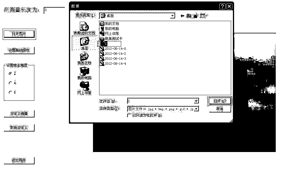
[0016] 3. Open the software written by me (software download address is drjcjjlq163.com password drjcjjlq1+1 or zlsq2012126.com password zlsqyx123). Click the Open Picture button to open the collected picture saved locally.

[0017] 4. Measure the pixel value between the jaws of the biopsy forceps. Click the left mouse button on one end of the jaws of the biopsy forceps, press and hold the left mouse button, drag the mouse to the other end ...


PUM


Abstract

The invention relates to the field of the medical endoscope (gastroscopy and colonoscopy) inspection technology, in particular to accurate measurement of the size of a nidus under an endoscope. An open jaw of measurable biopsy forceps is introduced as a reference substance.The length between the two ends of the open jaw of the biopsy forceps outside a body is measured in advance. The jaw of the biopsy forceps is placed on the same plane as the nidus. The distance from the plane where the jaw of the biopsy forceps is located to a lens of the endoscope is made to be equal to the distance from the plane where the nidus is located to the lens of the endoscope. The pixel value between the two ends of the jaw of the biopsy forceps under the endoscope and the pixel value of the radial line of the nidus are measured through software, and then the physical size of the radial line of the nidus can be calculated.

Description

technical field [0001] The invention is applied to the technical field of medical endoscopy (gastroscopy, colonoscopy), especially the measurement of the size of the lesion (ulcer, tumor) under the endoscope. Background technique [0002] The biological characteristics of the lesions (ulcers, tumors) that occur in the digestive tract are different in size. Now there are software measurement method and visual inspection method for the measurement methods of ulcers and tumors. The software measurement method is to measure with the software attached to the endoscopy workstation. Because it does not take into account the zoom effect of the endoscope on the measured object, the measurement result is inaccurate and unreliable. The visual method is to determine the size of the lesion by experienced endoscopists through visual inspection. This subjective result is even more inaccurate, and different people will give different results for the same lesion. Contents of the inventio...

Claims


Application Information

Patent Timeline
IPC IPC(8): A61B5/107
Inventor 吕海涛李英楠
Owner 吕海涛
Learn More