Method for establishing heart disease drug screening model

A heart disease and drug technology, applied in the field of biomedicine, can solve the problems of different effects and side effects, and achieve the effect of wide application prospects.

Inactive Publication Date: 2014-01-15
SHANGHAI BODE BIOTECH
View PDF0 Cites 3 Cited by
  • Summary
  • Abstract
  • Description
  • Claims
  • Application Information

AI Technical Summary

Problems solved by technology

At present, there are many types of drugs targeting beta adrenergic receptors clinically, but most of them have relatively large

Method used


Image

  • Method for establishing heart disease drug screening model
  • Method for establishing heart disease drug screening model
  • Method for establishing heart disease drug screening model

Examples


Embodiment 1

[0029] Embodiment 1, the preparation of IPS cell

[0030] 1.1. Four transcription factors, Oct3 / 4, Sox2, c-Myc and klf4, are coated with lentivirus

[0031] Four transcription factors, Oct3 / 4, Sox2, c-Myc and klf4, were amplified from total mouse cDNA by PCR. The primer sequences used in PCR amplification are as follows:

[0032] Oct3 / 4: 5' Primer: 5'-ATGGCTGGACACCTGGCTTCAGAC-3'

[0033] 3' Primer: 5'-GTTTGAATGCATGGGAGAGCCCAG-3'

[0034] Sox2: 5' Primer: 5'-ATGTATAACATGATGGAGACGGAG-3'

[0035] 3' Primer: 5'-CATGTGCGACAGGGGCAGTG-3'

[0036] c-Myc: 5' Primer: 5'-ATGCCCCTCAACGTGAACTTCAC-3'

[0037] 3' Primer: 5'-TGCACCAGAGTTTCGAAGCTGTTC-3'

[0038] klf4: 5' Primer: 5'-ATGAGGCAGCCACCTGGCG-3'

[0039] 3' Primer: 5'-AAAGTGCCTCTTCATGTGTAAGG-3'.

[0040] The PCR amplification products of the four transcription factors were respectively inserted into the lentiviral vector lenti-ef1a-EGFP (purchased from Invitrogen), and co-transfected into virus packaging cells, namely 293T cel...

Embodiment 2

[0050] Example 2, Identification of IPS cell totipotency

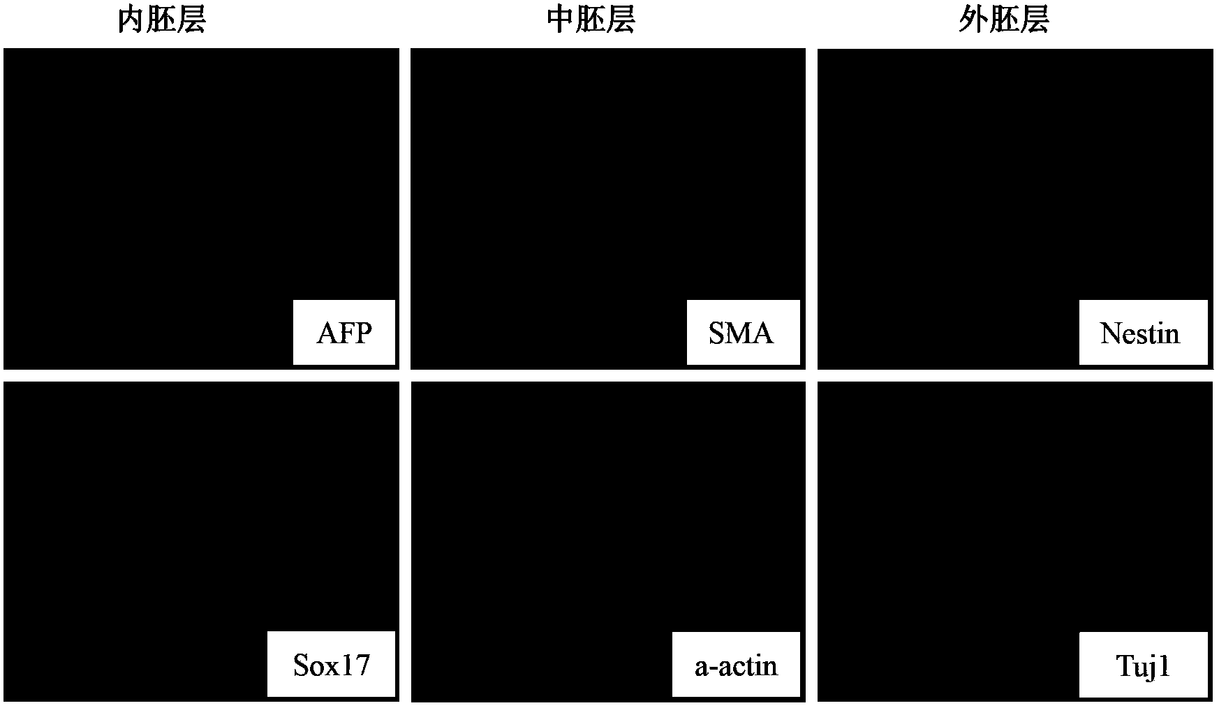
[0051] In order to verify the pluripotency of the IPS cells obtained in Example 1 and make them naturally differentiate to form embryoid bodies (EBs), the embryoid bodies were sliced ​​and stained with three germ layer-specific Marker antibodies to observe the embryoid bodies (EBs) of each germ layer. Express the situation. Among them, endoderm-specific markers are AFP and Sox17; mesoderm-specific markers are SMA and a-actin; ectoderm-specific markers are Nestin and Tuj1. The result is as figure 1 As shown, colony formation can be observed.

Embodiment 3

[0052] Example 3, IPS cells are directed to differentiate into cardiomyocytes

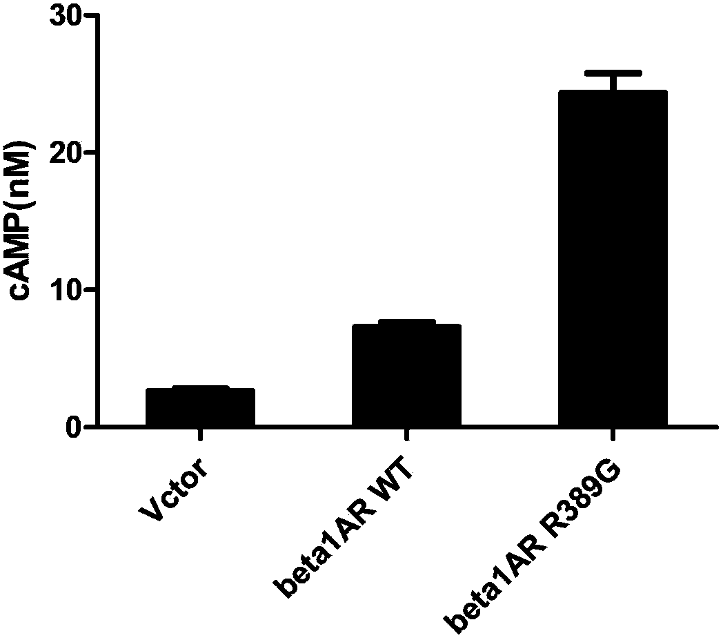
[0053] The wild-type beta1AR and beta2AR genes were mutated into beta1ARR389G and beta2AR G16R genes by site-directed mutagenesis, and then the wild-type beta1AR and beta2AR genes and mutant beta1AR R389G and beta2AR G16R genes were inserted into the lentiviral vector lenti-ef1a-EGFP ( purchased from Invitrogen), and co-transfected virus packaging cells, namely 293T cells (from Duke University, USA). Cultivate for 48-72hrs, and collect the supernatant culture solution containing the virus. Pass the supernatant through a 45 μm filter, then transfer to a sterile storage tube and save for later use.

[0054] The IPS cells obtained in Example 1 were infected with the above-mentioned lentivirus, and the experiment was divided into 3 groups:

[0055] a. Blank control group: lentivirus carrying green fluorescent protein;

[0056] b. Transfection of wild-type beta1AR and mutant beta1AR R389G into beta1A...


PUM


Abstract

The invention provides a method for establishing a heart disease drug screening model, which comprises the following steps of A) establishing a virus vector comprising four transcription factors: Oct3/4, Sox2, c-Myc and klf4; B) transfecting the virus vector in the step A) with a virus packaging cell to obtain virus liquid; C) infecting the virus liquid in the step B) with the fibroblast of the beta adrenergic receptor wild-type gene knockout mouse; D) screening the clone similar to the mouse embryonic stem cell, and subculturing to obtain the induced pluripotent stem cells with beta adrenergic receptor wild-type gene defect; E) establishing a virus vector containing the beta adrenergic receptor mutant gene; F) transfecting the virus vector in the step E) with the virus packaging cell to obtain virus liquid; G) infecting the virus liquid in the step F) with the induced pluripotent stem cells with beta adrenergic receptor wild-type gene defect; H) selecting positive clone and performing induced differentiation into myocardial cells.

Description

technical field [0001] The invention belongs to the field of biomedicine, in particular to a method for establishing a drug screening model for heart disease. Background technique [0002] The heart is a non-renewable organ in the human body. Heart disease cannot be repaired by itself and will have a fatal impact on patients. Therefore, the treatment and diagnosis of heart disease has become a medical problem. Stem cells are a type of special cells with self-renewal and pluripotent differentiation potential, which can differentiate into various types of cells in the human body. In 1981, the laboratories of Martin John Evans and Matthew Kaufman successfully cultured mouse embryonic stem cells (ES cells). A number of studies have shown that embryonic stem cells cultured in vitro can be induced to become cardiomyocytes. Therefore, the use of stem cells to differentiate into cardiomyocytes can provide new ideas for the treatment, diagnosis and drug evaluation of heart diseases....

Claims


Application Information

Patent Timeline
IPC IPC(8): C12N15/86C12N15/867C12N5/10C12Q1/02
Inventor 洪梁
Owner SHANGHAI BODE BIOTECH
Why Eureka
Try Eureka