
Tumor marker and application thereof to preparation of colorectal cancer diagnostic reagent

A technology of colorectal cancer and diagnostic reagents, applied in the field of serum/tissue tumor markers, can solve the problems of failure to reach diagnosis, limited value of early diagnosis of colon cancer, and inaccurate indicators of pancreatic cancer

Active Publication Date: 2014-04-23
THE THIRD AFFILIATED HOSPITAL OF SUN YAT SEN UNIV
View PDF3 Cites 15 Cited by
  • Summary
  • Abstract
  • Description
  • Claims
  • Application Information

AI Technical Summary

Problems solved by technology

However, these markers are imprecise and less sensitive as indicators for the diagnosis of pancreatic cancer alone, and they are also elevated in other gastrointestinal tumors and benign diseases, so the value of early diagnosis of colon cancer is limited
In addition, some genetic tumor markers such as p53, k-ras, etc. are also used in the early diagnosis of clinical colon cancer, but they have not been able to achieve the purpose of diagnosis

Method used


Image

  • Tumor marker and application thereof to preparation of colorectal cancer diagnostic reagent
  • Tumor marker and application thereof to preparation of colorectal cancer diagnostic reagent
  • Tumor marker and application thereof to preparation of colorectal cancer diagnostic reagent

Examples


Embodiment 1

[0048] Embodiment 1 sample collection and processing

[0049] Tissue samples from 7 patients with colon cancer and 10 samples from the control group were clinically collected, and the clinical data of the patients were systematically collected.

Embodiment 2

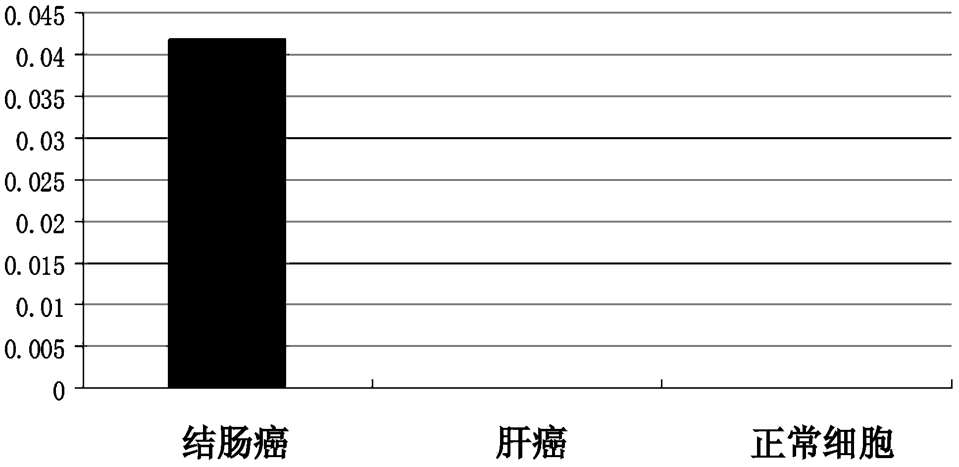
[0050] Detection of mRNA in embodiment two colon cancer and normal cells

[0051] 1. Cell total RNA extraction, use chloroform extraction method to extract the total RNA of cells, the specific steps are as follows:

[0052] (1) Take the colon cancer, liver cancer and normal cells in the medium dish, add 1ml Trizol reagent to lyse the tissue, repeatedly blow with a gun or shake vigorously to lyse the cells;

[0053] (2) Transfer the Trizol lysate of the above cells into an EP tube, and place it at room temperature 15-30°C for 5 minutes; (3) Add 0.2 ml of chloroform to the above EP tube according to the amount of 0.2 ml of chloroform per 1 ml of TRIZOL, and cover Put the cap on the EP tube, shake it vigorously in your hand for 15 seconds, let it stand at room temperature (15°C-30°C) for 2-3 minutes, then centrifuge at 12000g (4°C) for 30 minutes;

[0054] (4) After centrifugation, take the upper aqueous phase and put it in a new EP tube, add 1 volume of isopropanol according to...

Embodiment 3

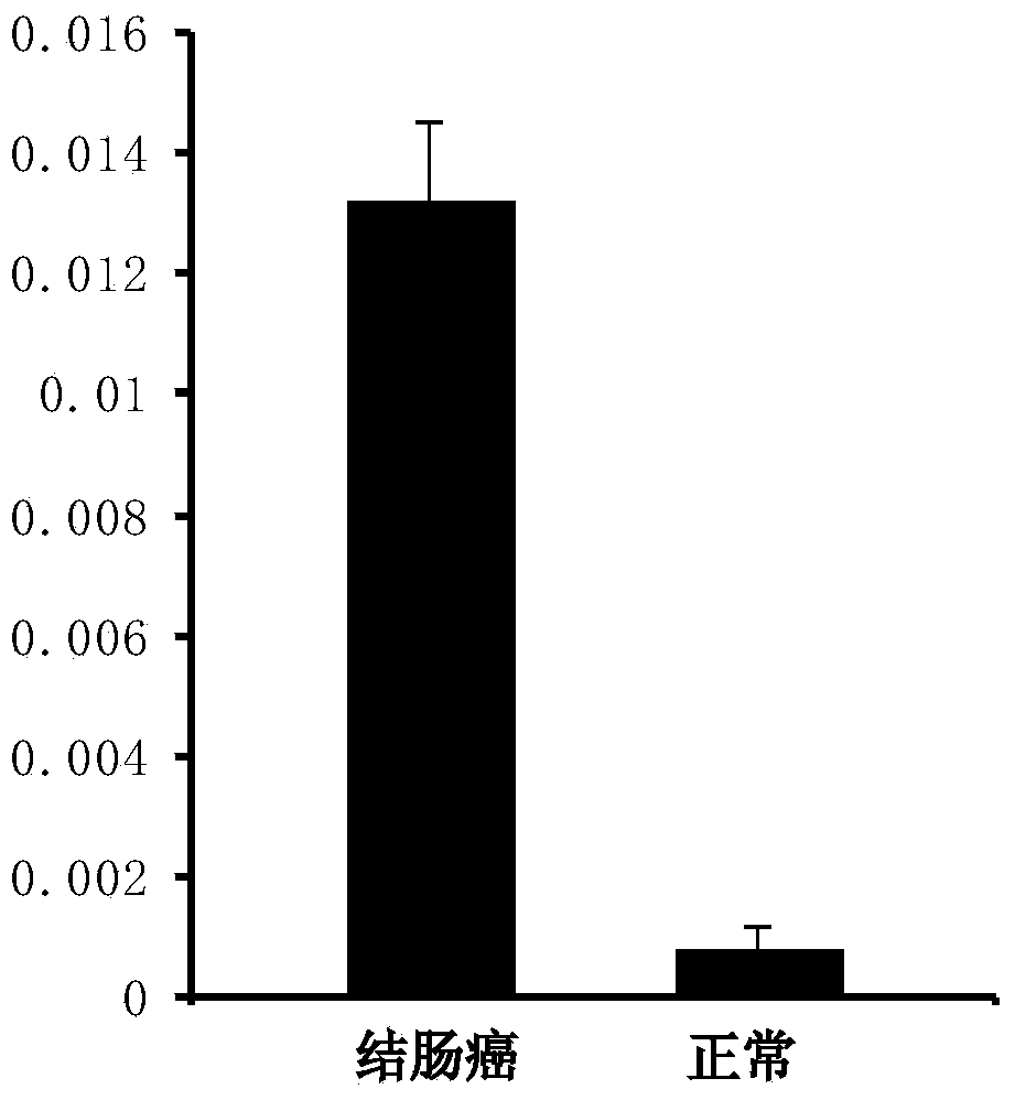
[0070] Example 3 CST1, CST4, CST2mRNA qRT-PCR experiments in tissues

[0071] 1. According to the cell results, CST1, CST4, and CST2 were further verified by qRT-PCR.

[0072] 2. Extraction of total RNA from tissue samples, using the chloroform extraction method to extract total RNA from tissue samples, the specific steps are as follows:

[0073] (9) Take the patient's frozen colon tissue sample, knock out a tissue block with a size of about 2mm×2mm×2mm, add 1ml Trizol reagent to lyse the tissue, and repeatedly use a gun to blow or shake vigorously to lyse the tissue; (10) The above tissue Transfer the Trizol lysate into an EP tube and place it at room temperature 15-30°C for 5 minutes;

[0074] (11) In the above EP tube, add 0.2ml chloroform for every 1ml TRIZOL, cover the EP tube cap, vibrate vigorously in your hand for 15 seconds, and place it at room temperature (15°C-30°C) for 2-3 minutes Afterwards, centrifuge at 12000g (4°C) for 30 minutes;

[0075] (12) After centri...


PUM


Abstract

The invention belongs to the field of genetic engineering and medicines, and relates to a novel tumor marker for early diagnosis of colorectal cancer, in particular to a tumor marker related to the colorectal cancer and application thereof. The marker is a composition of CystatinSN (CST1), CystatinS (CST4) and CystatinSA (CST2). The marker as well as a primer and an antibody thereof can be applied to a diagnostic kit for the auxiliary diagnosis of the colorectal cancer.

Description

technical field [0001] The invention belongs to the fields of genetic engineering and oncology, and relates to a serum / tissue tumor marker related to colorectal cancer and its application. Background technique [0002] Colon cancer and rectal cancer are common malignant tumors, most of them are advanced, and their incidence rate ranks fourth among malignant tumors. With economic development, changes in lifestyle, living environment and dietary structure, the incidence of colorectal cancer is on the rise. Although the diagnostic level of colorectal cancer has improved, the detection rate of early colorectal cancer is still low and the mortality rate is high. According to statistics, the 5-year survival rate of early colon cancer after radical surgery is about 80%, while the 5-year survival rate of advanced colon cancer is only 50%. Therefore, early diagnosis is a key factor in improving the cure rate of colon cancer, and finding tumor markers with high sensitivity and speci...

Claims


Application Information

Patent Timeline
IPC IPC(8): C12Q1/68C12N15/11C07K14/81G01N33/68G01N33/574
CPCC12Q1/6886C12Q2600/158G01N33/57419G01N33/57446
Inventor 吴斌
Owner THE THIRD AFFILIATED HOSPITAL OF SUN YAT SEN UNIV
Learn More