Full-eye optical coherence tomography imager

An optical coherence tomography and coherence tomography technology, which is applied in the fields of ophthalmoscope, medical science, eye testing equipment, etc. The effect of small image distortion

Active Publication Date: 2014-05-28
INST OF OPTICS & ELECTRONICS - CHINESE ACAD OF SCI
View PDF6 Cites 28 Cited by
  • Summary
  • Abstract
  • Description
  • Claims
  • Application Information

AI Technical Summary

Problems solved by technology

The system has the following disadvantages: 1) The light source is the most expensive device in the OCT system, and the use of two light sources will greatly increase the cost; 2) The dichroic prism used in the sample arm halves the intensity of the illumination light signal from the light source, The intensity of the light signal returned from the sample will be halved again, and the extremely low utilization rate of light energy will reduce the detection sensitivity of the system; 3) Much of the information of the anterior segment is distributed on the nasal side and the temporal side, requiring large-scale scanning and imaging, while the fundus The imaging field of view is extremely limited, and a common set of scanning mechanisms will not be able to perform scanning settings separately according to the requirements of the respective field of view.
The structure of this system is greatly simplified, but there are also shortcomings: 1) The imaging optical path of the anterior segment and the optical path of the reference arm are seriously mismatched, making it difficult to obtain a clear image of the anterior segment; It has a very long coherence length, which means that the light source is required to have a very high wavelength scanning resolution, which brings difficulties to the production of the light source; 2) cannot eliminate the conjugate artifacts of the anterior segment and fundus at the same time; 3) share a set of scanning Mechanisms, scanning settings cannot be made separately according to the requirements of the respective fields of view

Method used


Image

  • Full-eye optical coherence tomography imager
  • Full-eye optical coherence tomography imager

Examples


Embodiment Construction

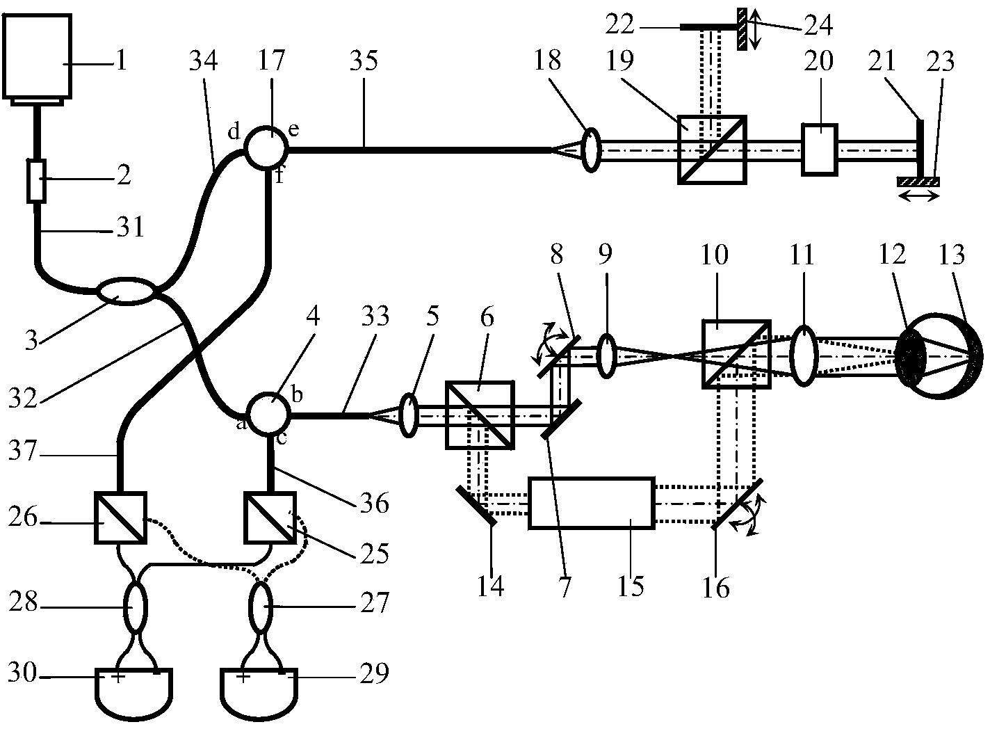
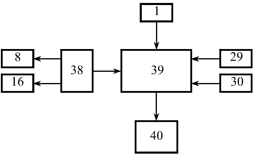
[0026] The structure of the whole-eye optical coherence tomography imager proposed by the present invention is as follows: figure 1Shown, including: frequency-sweeping light source 1, fiber polarizer 2, first polarization-maintaining optical coupler 3, first optical circulator 4, first lens 5, first broadband polarization beam splitter prism 6, first reflector 7, the first A two-dimensional scanner 8, a second lens 9, a second broadband polarization beam splitter prism 10, a third lens 11, a second mirror 14, a beam expander 15, a second two-dimensional scanner 16, and a second optical circulator 17 , the fourth lens 18, the third broadband polarization beam splitter prism 19, water box 20, first and second reference mirrors 21-22, first and second translation stage 23-24, the fourth and the fifth broadband polarization beam splitter prism 25 -26, second and third polarization-maintaining optical couplers 27-28, first and second balanced detectors 29-30, first to seventh singl...


PUM


Abstract

A full-eye optical coherence tomography imager comprises a frequency sweeping light source, an optical fiber polarizer, a first optical circulator, a second optical circulator, a first broadband polarization splitting prism, a second broadband polarization splitting prism, an ocular anterior segment scanning imaging light path, an eye ground scanning imaging light path, a third broadband polarization splitting prism, two reference mirrors, a fourth broadband polarization splitting prism, a fifth broadband polarization splitting prism, a second polarized light protecting coupler, a third polarized light protecting coupler, a first balance detector, a second balance detector, a function generation card, a data acquisition card, a computer and the like, wherein the first broadband polarization splitting prism, the second broadband polarization splitting prism, the ocular anterior segment scanning imaging light path and the eye ground scanning imaging light path are arranged in a sample arm; the third broadband polarization splitting prism and the two reference mirrors are arranged in a reference arm; the fourth broadband polarization splitting prism, the fifth broadband polarization splitting prism, the second polarized light protecting coupler, the third polarized light protecting coupler, the first balance detector and the second balance detector are arranged at a detecting end. The imager is based on a frequency sweeping optical coherence tomography technology, a component p and a component s of a polarized beam are used for conducting imaging on an eye ground and an ocular anterior segment respectively, and thus three-dimensional high resolution real-time imaging can be conducted on an full eye structure at the same time on the same system without any device for conversion. By means of the full-eye optical coherence tomography imager, optical path length and dispersion matching and scan setting can respectively be carried out on ocular anterior segment imaging and eye ground imaging, and thus the optimal imaging quality and the view field size of ocular anterior segment imaging and eye ground imaging can be obtained at the same time.

Description

technical field [0001] The invention relates to human eye imaging equipment and optical coherence tomography (OCT) technology, in particular to an equipment that adopts frequency-sweeping OCT technology and can simultaneously image the whole eye structure. Background technique [0002] Eye diseases such as short-sightedness, long-sightedness, cataracts and glaucoma can affect the shape and size of the eye. Simultaneous high-resolution imaging of whole-ocular structures is essential in order to examine changes in visual physiology due to ophthalmic diseases. Ultrasound and MRI techniques can achieve whole-eye imaging, but the former is a contact measurement, while the latter is expensive, and their resolution is too low to effectively observe tissue structures. Optical coherence tomography (OCT) technology is a technology that can perform three-dimensional high-resolution non-destructive real-time tomographic imaging of the internal structure of tissues. widely used. At pr...

Claims


Application Information

Patent Timeline
Patent Type & Authority Applications(China)
IPC IPC(8): A61B3/14A61B3/12
Inventor 杨亚良张雨东饶学军王成
Owner INST OF OPTICS & ELECTRONICS - CHINESE ACAD OF SCI
Why Eureka
Try Eureka