Renal artery blood-supply area segmenting method based on CT contrastographic picture

A technology of contrast image and region segmentation, which is applied in the field of image processing technology, can solve the problems of affecting the operation effect, inaccurate estimation, and large variation of renal artery anatomical structure, so as to improve the reliability and improve the effect of operation

Inactive Publication Date: 2014-06-11
SOUTHEAST UNIV
View PDF4 Cites 12 Cited by
  • Summary
  • Abstract
  • Description
  • Claims
  • Application Information

AI Technical Summary

Problems solved by technology

However, due to the great variability in the anatomy of the renal arteries (e.g. figure 2 As shown, the picture is taken from the paper "Morphological assessments on the arteries of the superior renal segment", Surgical and Radiologic Anatomy, 2012,34(2):pages137-144) published by B.Petru et al., only based on visual observation and experience Judgment often leads to inaccurate estimation, affects the operation effect or causes operation failure

Method used


Image

  • Renal artery blood-supply area segmenting method based on CT contrastographic picture
  • Renal artery blood-supply area segmenting method based on CT contrastographic picture
  • Renal artery blood-supply area segmenting method based on CT contrastographic picture

Examples


Embodiment Construction

[0022] The present invention will be further described below in conjunction with the accompanying drawings.

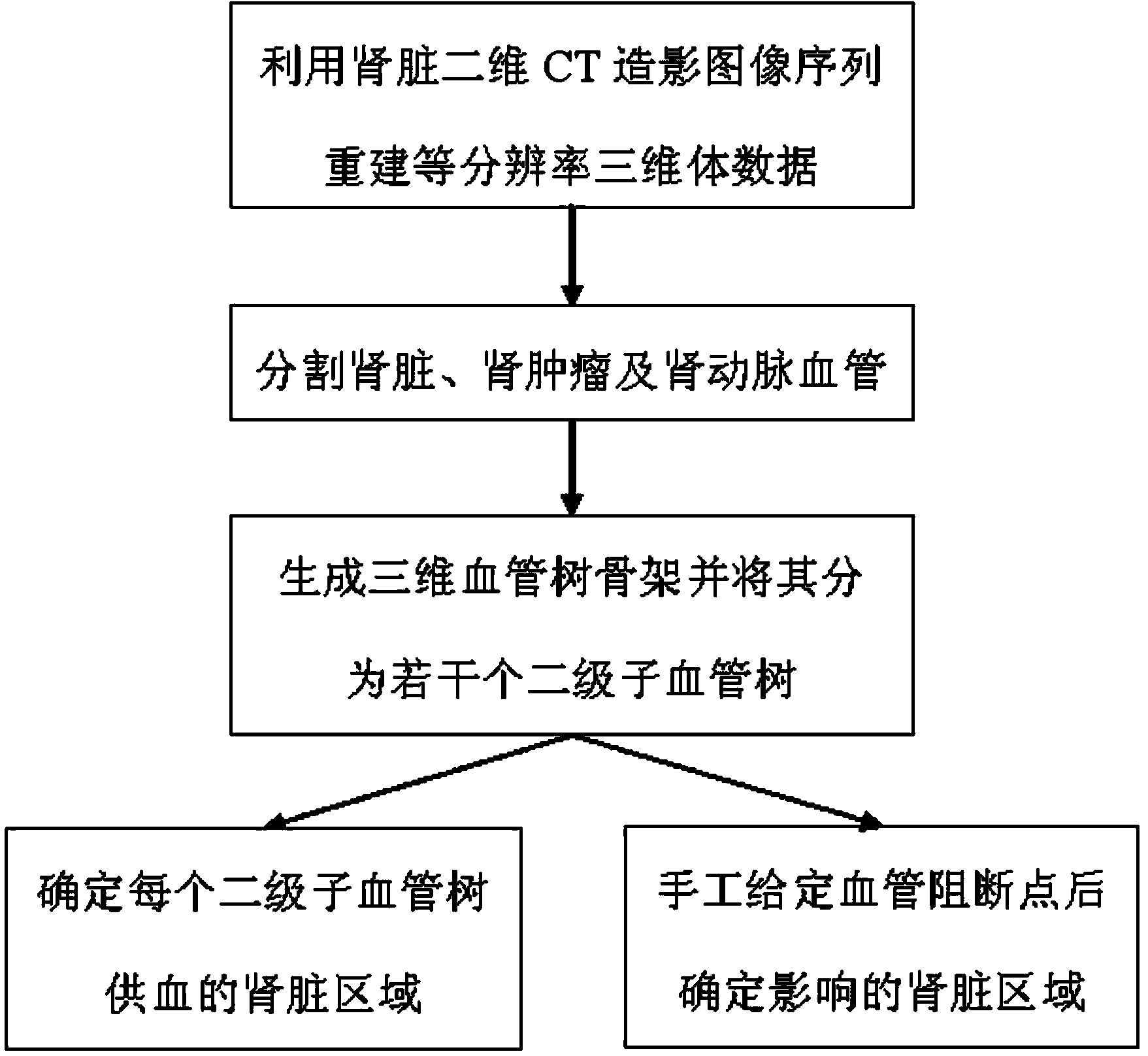
[0023] Such as image 3 Shown is a method for segmenting the blood supply area of ​​the renal artery based on the CT contrast image, using the three-dimensional CT contrast data of the kidney to determine the blood supply range of each branch of the renal artery, specifically including the following steps executed in sequence:

[0024] (1) Using the interpolation algorithm to reconstruct the two-dimensional CT contrast image sequence into three-dimensional volume data with equal resolution in all directions;

[0025] (2) In the three-dimensional volume data, the kidney, renal tumor area, and renal artery blood vessel were segmented respectively, and the segmentation results were represented by binarized volume data; specifically:

[0026] (2.1) Segmentation of the kidney area: the paper "Automatic Detection and Segmentation of Kidneys in 3D CT Images Using Random Fore...


PUM


Abstract

The invention discloses a renal artery blood-supply area segmenting method based on a CT contrastographic picture. The method comprises the steps that firstly, the kidneys, kidney neoplasms and the renal arteries in the CT contrastographic picture are segmented, then segmented kidney tissue areas are calculated to obtain Euclidean distances of different renal artery branches, and a blood-supply area of each renal artery secondary blood vessel tree is determined according to the minimum Euclidean distance. The method not only can be used for accurately estimating kidney neoplasm blood supply branches, but also can simulate the kidney blood-supply areas possibly affected when any position of the renal artery branches is blocked. As the renal artery anatomical structures of different patients have large differences, according to the method, the blood-supply areas of the renal artery branches can be accurately determined through the CT contrastographic picture of the kidneys, the more visual and reliable basis is provided for a pre-operation project of a partial kidney resection operation, and the treatment effect and the success rate are improved.

Description

technical field [0001] The invention relates to a method for segmenting renal artery blood supply area based on CT contrast images, and relates to image processing technology. Background technique [0002] Renal cancer is a common malignant tumor that occurs in the kidney. In the past 20 years, the incidence of renal tumors in my country has increased at an average rate of 6.5% per year, ranking first among urological tumor-related deaths. In the past, the method of directly removing the entire diseased kidney was usually used in the treatment of renal cancer. Although this method can remove the entire lesion area, it also greatly affects the quality of life of the patient after surgery. At present, it is internationally recognized that the tumor size is less than 4 cm. There is no difference in tumor control between total nephrectomy and partial nephrectomy. Partial resection can allow patients to retain more renal function and is more beneficial to patient survival after s...

Claims


Application Information

Patent Timeline
Patent Type & Authority Applications(China)
IPC IPC(8): G06T7/00
Inventor 杨冠羽王征唐立钧沈傲东舒华忠
Owner SOUTHEAST UNIV
Why Eureka
Try Eureka