Phase microscope imaging method based on SHWS (Shack-Hartmann Wavefront Sensor)

A phase microscope and imaging method technology, applied in the field of microscope imaging, can solve the problems of cumbersome reconstruction process, vibration noise, complex hardware structure and so on

Active Publication Date: 2014-06-25
ZHEJIANG UNIV
View PDF9 Cites 3 Cited by
  • Summary
  • Abstract
  • Description
  • Claims
  • Application Information

AI Technical Summary

Problems solved by technology

[0007] Aiming at the above-mentioned technical problems existing in the prior art, the present invention provides a phase microscope imaging ...

Method used


Image

  • Phase microscope imaging method based on SHWS (Shack-Hartmann Wavefront Sensor)
  • Phase microscope imaging method based on SHWS (Shack-Hartmann Wavefront Sensor)
  • Phase microscope imaging method based on SHWS (Shack-Hartmann Wavefront Sensor)

Examples


Embodiment Construction

[0029] In order to describe the present invention more specifically, the technical solutions of the present invention will be described in detail below in conjunction with the accompanying drawings and specific embodiments.

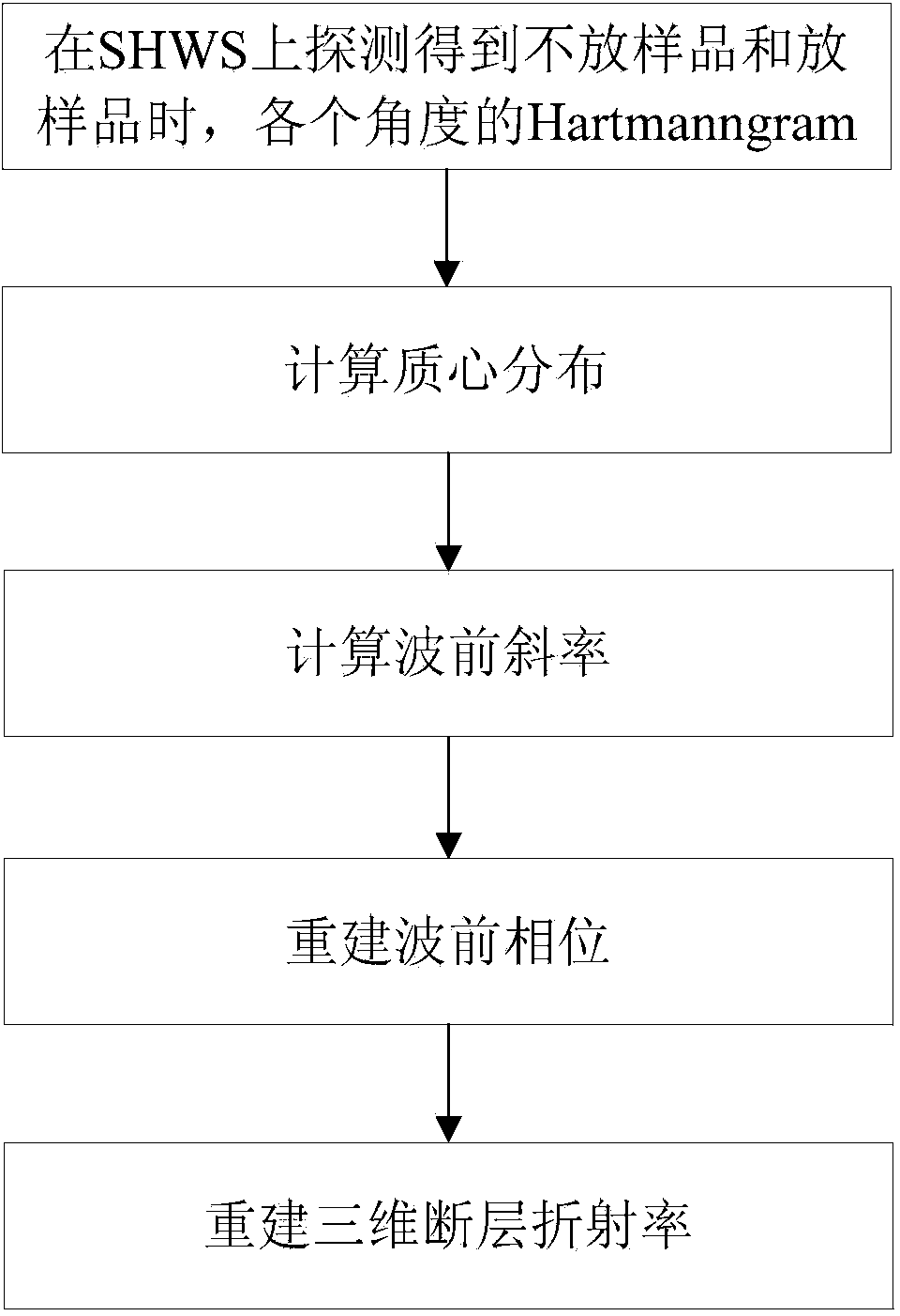
[0030] Such as figure 1 Shown, a phase microscope imaging method based on SHWS, comprises the following steps:

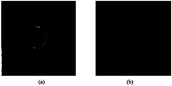
[0031] (1) For the two cases of not putting the sample and putting the sample, use SHWS to detect and obtain the Hartmanngram corresponding to the light at each angle in the two cases;

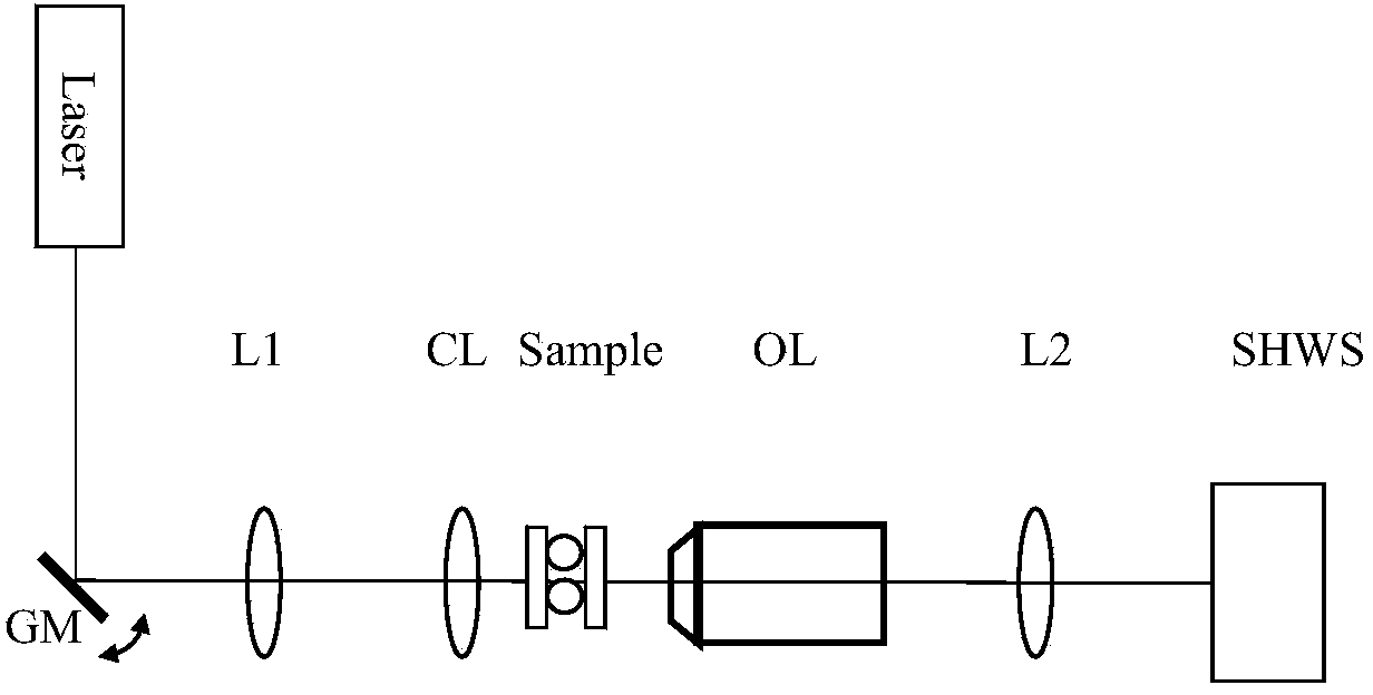
[0032] Such as figure 2As shown, the helium-neon laser beam He-Ne (λ=633nm) is first incident on the scanning galvanometer GM. Then, after passing through lens L1 (f=200mm) and oil-immersed condenser lens CL (Nikon, NA=1.4) in turn, it is irradiated onto the sample. A 4f optical system is formed between the GM and the sample. The sample is immersed in a solution with a certain refractive index and placed between two coverslips, which are separated and seal...


PUM


Abstract

The invention discloses a phase microscope imaging method based on an SHWS (Shack-Hartmann Wavefront Sensor). The phase microscope imaging method comprises the following steps: (1) collecting Hartmanngram; (2) calculating mass center distribution; (3) calculating wavefront slope; (4) reconstructing a wavefront phase; and (5) reconstructing a three-dimensional fault refractive index. According to the phase microscope imaging method, the problems of a phase microscope based on an interference method that the establishment of hardware is complicated, and a reconstruction step is complicated and a lot of noises are easily caused by vibration influence can be improved by effectively utilizing the SHWS; compared with an experiment of an existing reconstruction method, the experiment of the method according to the invention has the advantages that the hardware is greatly simplified, the reconstruction step is simplified and the vibration influences are avoided.

Description

technical field [0001] The invention belongs to the technical field of microscope imaging, and in particular relates to a phase microscope imaging method based on SHWS (Shack-Hartmann Wavefront Sensor, Shack-Hartmann Wavefront Sensor). Background technique [0002] In recent years, with the advancement of science and technology, optical microscopy has become a research hotspot for biologists and clinical researchers with its higher spatial resolution, greater imaging depth and the feasibility of non-destructive detection of living cells. It has become an indispensable tool in biology and medical research, and is an important basis for the establishment and development of cytology and cell biology. At present, in cell biology, in addition to ordinary optical microscopes, there are many kinds of microscopes such as fluorescence microscopes, laser confocal microscopes, and phase microscopes. [0003] Among them, phase microscopy techniques include phase contrast microscopy, di...

Claims


Application Information

Patent Timeline
IPC IPC(8): G01N21/41G02B21/00
Inventor 刘华锋黄晨曦
Owner ZHEJIANG UNIV
Why Eureka
Try Eureka