Dynamic brain functional magnetic resonance imaging method and system

A magnetic resonance imaging and brain function technology, applied in medical science, sensors, diagnostic recording/measurement, etc., can solve the problem of insufficient temporal and spatial resolution of imaging, and achieve the effect of improving the quality of reconstructed images and improving temporal and spatial resolution

Inactive Publication Date: 2014-07-23
UNIV OF SCI & TECH OF CHINA
View PDF3 Cites 10 Cited by
  • Summary
  • Abstract
  • Description
  • Claims
  • Application Information

AI Technical Summary

Problems solved by technology

Although a lot of research has been devoted to improving the temporal or spatial resolution of fMRI imaging in rece...

Method used


Image

  • Dynamic brain functional magnetic resonance imaging method and system
  • Dynamic brain functional magnetic resonance imaging method and system
  • Dynamic brain functional magnetic resonance imaging method and system

Examples


Embodiment 1

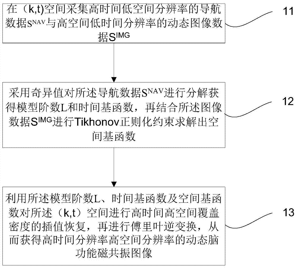
[0025] figure 1 It is a flow chart of a dynamic brain functional magnetic resonance imaging method provided in Embodiment 1 of the present invention. Such as figure 1 As shown, the method mainly includes the following steps:

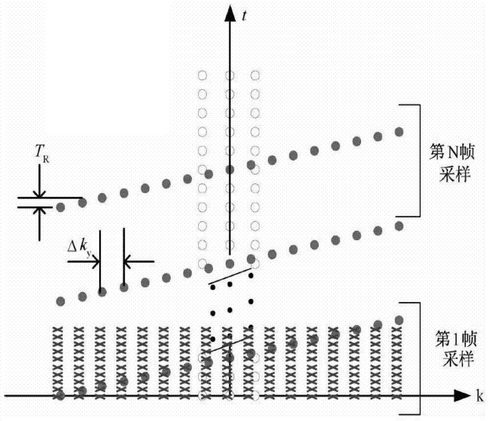
[0026] Step 11. Collect navigation data S with high time and low spatial resolution in (k, t) space NAV Dynamic image data with high spatial and low temporal resolution S IMG .

[0027] In the (k, t) space, each moment along the time axis t is a complete k-space, where k is a two-dimensional space.

[0028] In the embodiment of the present invention, based on the preset number of phase encoding steps and the echo chain length (ETL), the corresponding number of excitations is generated; wherein, the number of K-space phase encoding lines is filled after each radio frequency pulse excitation, and in the same radio frequency cycle The navigation data SNAV and the image data SIMG are collected synchronously.

[0029] Step 12, using the singular value f...

Embodiment 2

[0045] In order to facilitate understanding of the present invention, below in conjunction with Figure 2-5 Make further introductions.

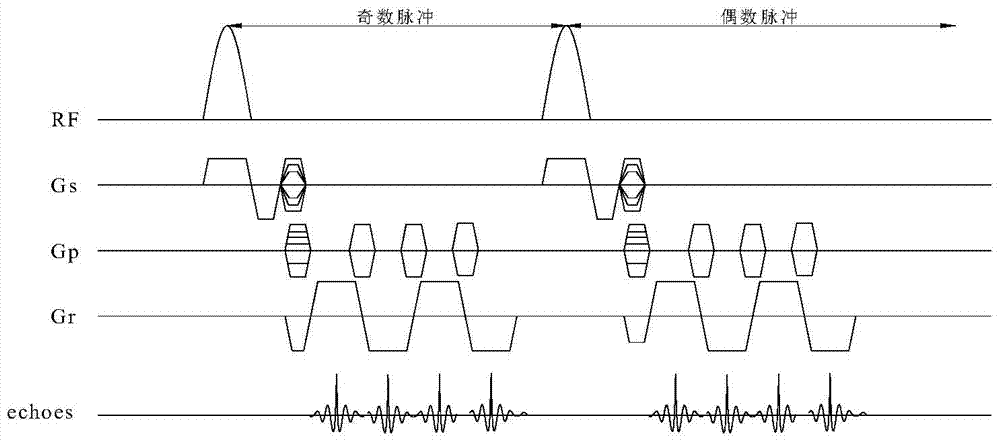
[0046] The embodiment of the present invention provides a dynamic brain functional magnetic resonance imaging method, which fully exploits the advantages of the PSF model and the MS (multiple excitation echo planar imaging technology)-EPI sequence, and solves the partially separable function model using Tikhonov regular constraints The key parameters in the method can improve the quality of the reconstructed image while releasing the constraints of time and space.

[0047] Among them, brain functional imaging sequence (EPI) is developed on the basis of gradient echo, and can be divided into single-shot echo planar imaging (SS-EPI) and multiple-shot echo planar imaging according to the number of excitations. Technology (multi-shot EPI, MS-EPI). The raw data of all K-spaces can be obtained with a single shot, and the imaging time is signific...

Embodiment 3

[0078] Figure 6 It is a schematic diagram of a dynamic brain functional magnetic resonance imaging system provided by Embodiment 3 of the present invention. Such as Figure 6 As shown, the system mainly includes:

[0079] The data collection module 61 is used to collect navigation data S with high time and low spatial resolution in (k, t) space NAV Dynamic image data with high spatial and low temporal resolution S IMG ;

[0080] Calculation module 62, for using the singular value to calculate the navigation data S NAV Decompose to obtain the model order L and time basis function Combined with the image data S IMG Perform Tikhonov regularization constraints to solve the spatial basis function c l (k);

[0081] Imaging module 63, for using the model order L, time basis function and spatial basis functions c l (k) performing interpolation recovery of high temporal and high spatial coverage density on the (k, t) space, and then performing inverse Fourier transform, so...


PUM


Abstract

The invention discloses a dynamic brain functional magnetic resonance imaging method and system. The method includes the steps that high-time low-space-resolution navigation data S<NAV> and high-space low-time-resolution dynamic image data S are collected in (k,t) space; the navigation data S<NAV> are decomposed through singular values to obtain a model order L and a time primary function phi[l](t), and Tikhonov regularization constraint is conducted by combining with the image data S to solve a space primary function cl(k); interpolation recovery with the high-time high-space coverage density is conducted on the (k,t) space through the model order L, the time primary function phi[l](t) and the space primary function cl(k), Fourier inversion is then conducted, and therefore a high-time-resolution and high-space-resolution dynamic brain function magnetic resonance image is obtained. By means of the method and the system, quality of the reconstructed image is improved, and meanwhile the high-time-resolution and high-space-resolution dynamic brain function magnetic resonance image is obtained.

Description

technical field [0001] The invention relates to the technical field of magnetic resonance imaging, in particular to a dynamic brain functional magnetic resonance imaging method and system. Background technique [0002] Functional Magnetic Resonance Imaging (fMRI) is one of the fastest-growing fields in which contemporary medical imaging technology is applied to brain neuroscience research. The principle is to use magnetic resonance imaging to measure hemodynamic changes caused by neuronal activity. At present, it is mainly used in the study of the brain or spinal cord of humans and animals. Can show small changes in magnetic resonance signals arising from blood oxygenation status in venous capillaries in various regions of the brain. As a non-destructive detection technology, fMRI has become an important method to observe brain activity and reveal the relationship between brain and thinking, and it is used in almost every field of neuroscience, such as: neurophysiology, ne...

Claims


Application Information

Patent Timeline
IPC IPC(8): A61B5/055
Inventor 张晓咏李超邱本胜
Owner UNIV OF SCI & TECH OF CHINA
Why Eureka
Try Eureka