Fixed-decalcifying fluid for bone marrow biopsy and paraffin section method of bone marrow biopsy tissue

A technique of biopsy tissue and paraffin section, which is applied in the field of fixation-decalcification solution for bone marrow biopsy and paraffin section of bone marrow biopsy tissue, which can solve the problems of bone marrow biopsy that are not suitable for third-party independent laboratories, and shorten the preparation of specimens. Process, organizational structure is clear, well-preserved effect

Inactive Publication Date: 2014-08-20
WUHAN ADICON CLINICAL LAB
View PDF7 Cites 4 Cited by
  • Summary
  • Abstract
  • Description
  • Claims
  • Application Information

AI Technical Summary

Problems solved by technology

The existing bone marrow biopsy method needs to change the working solution midway, so the existing method is not suitable for third-party independent laboratories to complete the bone marrow biopsy project

Method used


Image

  • Fixed-decalcifying fluid for bone marrow biopsy and paraffin section method of bone marrow biopsy tissue
  • Fixed-decalcifying fluid for bone marrow biopsy and paraffin section method of bone marrow biopsy tissue
  • Fixed-decalcifying fluid for bone marrow biopsy and paraffin section method of bone marrow biopsy tissue

Examples


Embodiment 1

[0044] Example 1 Bone marrow biopsy tissue fixation-preparation of decalcification solution

[0045] Based on 1000ml, including

[0046] 0.1M Phosphate Buffer Saline (PBS) 900ml;

[0047] 40% formaldehyde 100ml;

[0048] EDTA 150 g;

[0049] Adjust the pH to 7.0-7.2.

[0050] Wherein, in addition to PBS, other suitable buffers can also be selected as the buffer.

Embodiment 2

[0051] Example 2 Bone marrow biopsy fixation-decalcification method

[0052] The bone marrow biopsy specimen was fixed and decalcified with the fixation-decalcification solution described in Example 1, wherein the volume ratio of sample:fixation-decalcification solution was equal to or greater than 1:10. Fix at room temperature - decalcify for 20-72 hours, then rinse with running water. The fixation and decalcification of the bone marrow tissue in the present invention are accomplished simultaneously in the fixation-decalcification solution.

Embodiment 3

[0053] Example 3 Method for Paraffin Section of Bone Marrow Biopsy

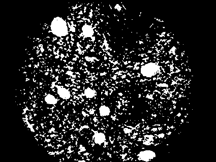
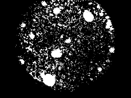
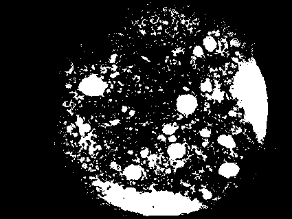
[0054] The specimens of bone marrow biopsy were fixed and decalcified with the fixation-decalcification solution and method described in Example 1 and Example 2 for 20-72 hours, rinsed with running water, dehydrated, soaked in wax, embedded, and sliced ​​(section thickness 2-3 μM ), HE staining and / or immunohistochemical staining, and observed under a microscope. The obtained slices can be directly used for microscope observation and pathological examination, and immunohistochemical staining can also be performed according to different needs. The obtained HE section tissue cells are well preserved and the staining effect is good. The positive immunohistochemical staining was accurately localized, and the background staining was light.

[0055] The dehydration method includes: putting the fixed decalcified bone marrow tissue into formaldehyde for 60 minutes, water for 10 minutes, 75% ethanol for 60 minutes, ...


PUM


Abstract

The present invention provides a fixed-decalcifying fluid for bone marrow biopsy and a paraffin section method of bone marrow biopsy tissue. The fixed-decalcifying fluid includes 40% of formaldehyde, EDTA and a buffer solution, and has a pH value of 7.0-7.2. The reagent and the method provided by the invention merge and complete the two steps of bone marrow tissue fixation and decalcification at the same time, don not require liquid change in the middle, reduce the operation steps, shorten the process of specimen pretreatment, and are more conducive to giving diagnosis conclusion and treatment by the doctor. The obtained HE section shows well preserved cell morphology and clear tissue structure; immunohistochemistry staining shows excellent staining effect, accurate positioning and clean background; and the sample does not show the phenomenon of excessive decalcification.

Description

technical field [0001] The invention relates to a fixation-decalcification solution for bone marrow biopsy and a method for paraffin sectioning bone marrow biopsy tissue. Background technique [0002] Bone marrow biopsy, referred to as bone marrow biopsy, is to use a special puncture needle to take a small piece of cylindrical bone marrow tissue about 1.0-2.0 cm long for pathological examination. The operation method is exactly the same as that of bone marrow smear aspiration. Bone marrow biopsy maintains a complete bone marrow tissue structure, which can well reflect the degree of bone marrow hyperplasia and provide a comprehensive understanding of the pathological characteristics of bone marrow, especially the bone marrow infiltration of extramedullary tumors. [0003] Paraffin section technique is now often used to prepare bone marrow biopsy tissue, which mainly includes the following steps: fixation of bone marrow sample tissue, decalcification, dehydration, embedding, s...

Claims


Application Information

Patent Timeline
Patent Type & Authority Applications(China)
IPC IPC(8): G01N1/36G01N1/28G01N1/30
Inventor 夏成青陈红梅
Owner WUHAN ADICON CLINICAL LAB
Why Eureka
Try Eureka