
A Visual Ventricle and Subdural Epidural Drainage System

A technology of external drainage and ventricle, which is applied in the field of medical devices, can solve problems such as inability to place the drainage tube in the best position, unsuccessful drainage, and exacerbation of the patient's condition, so as to avoid brain tissue damage or cerebral hemorrhage, and improve accuracy and efficiency. Improved effect

Active Publication Date: 2016-03-30
葛建伟
View PDF6 Cites 1 Cited by
  • Summary
  • Abstract
  • Description
  • Claims
  • Application Information

AI Technical Summary

Problems solved by technology

The existing external ventricular drainage tube insertion only depends on the doctor's clinical experience, and the drainage tube cannot be placed in the best position accurately, and the drainage may not be successful due to inaccurate placement; in addition, frequent brain punctures may lead to ventricular catheter insertion. Cerebral hemorrhage or ventricular hemorrhage will cause further increase in intracranial cerebral pressure (ICP) and aggravate the patient's condition
[0003] If the ventricular catheter is blocked by intraventricular choroid plexus tissue, blood clots, fragments of brain tissue, or fibrin debris, the external drainage may be poor and the operation may fail

Method used


Image

  • A Visual Ventricle and Subdural Epidural Drainage System
  • A Visual Ventricle and Subdural Epidural Drainage System
  • A Visual Ventricle and Subdural Epidural Drainage System

Examples


Embodiment Construction

[0038] The present invention will be specifically introduced below in conjunction with the accompanying drawings and specific embodiments.

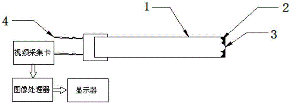
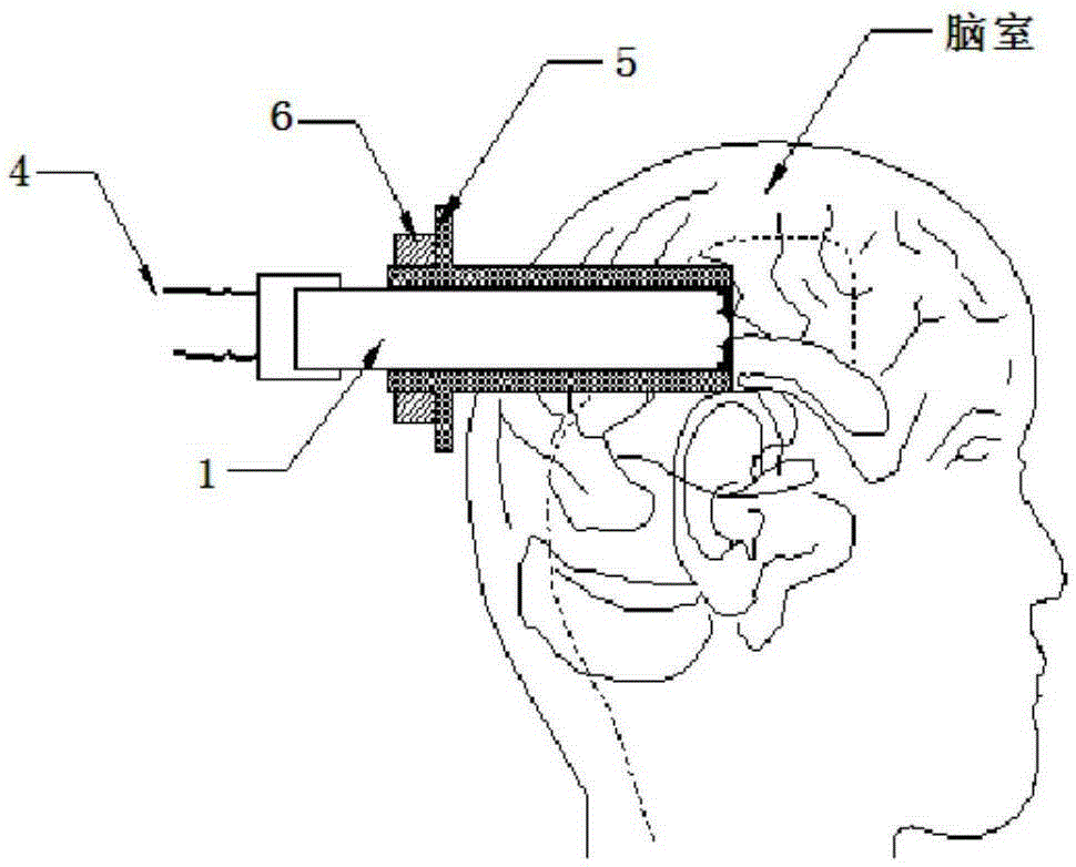
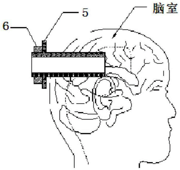
[0039] combine figure 1 , the visible ventricle and subdural extradural drainage system of the present invention includes a visible ventricle puncture system, and the visible ventricle puncture system includes a visible puncture guide core 1, and the intracranial end of the visible puncture guide core 1 is coaxial An LED light source 2 and a miniature camera 3 are provided, and the extracranial end is coaxially provided with a wire connected to a 5V DC power supply 4 and an output line of a video capture card, the output line of the video capture card is connected to the video capture card, and the output of the video capture card is Connect the image processor, the output of the image processor is connected to the display; combine figure 2 , the visible puncture guide core 1 can be connected and fixed with the puncture sheath 5 through...


PUM


Abstract

The invention discloses a visible cerebral ventricle and subdural external drainage system. The visible cerebral ventricle and subdural external drainage system comprises a visible cerebral ventricle puncture system and a drainage system. The visible cerebral ventricle puncture system comprises a visible puncture guiding core, and LED light sources and a miniature camera are coaxially arranged at the intracranial end of the visible puncture guiding core. The wall of a puncture sheath is see-through, and millimeter scales are arranged on the outer wall of the puncture sheath. The visible cerebral ventricle and subdural external drainage system has the remarkable advantages that as the miniature camera can be connected to one end of the visible puncture guiding core, and a monitor is connected to the other end of the visible puncture guiding core, cerebral puncture is completely performed in a direct-viewing mode, precision is improved, and it is avoided that due to operation based on a rule of thumb, the cerebral tissue is damaged, or cerebral hemorrhage occurs. As miniature vanes (fixing clamps) are arranged at the position distant from the cerebral ventricle by 10 cm-15 cm and used for fixing a drainage tube at the incised skin, the defect that a drainage tube used for the current clinic is simply bound and fixed through threads is overcome.

Description

technical field [0001] The invention relates to the technical field of medical devices, in particular to a visual ventricle and subdural external drainage system. Background technique [0002] External ventricular drainage or subdural hematoma drainage is an important surgical method for rescuing severe intracranial hemorrhage, obstructive hydrocephalus and intracranial hypertension. The existing external ventricular drainage tube insertion only relies on the doctor’s clinical experience, and the drainage tube cannot be placed in the best position accurately, and the drainage may not be successful due to inaccurate placement; in addition, frequent brain punctures may lead to ventricular catheter insertion. Cause cerebral hemorrhage or ventricular hemorrhage, and then cause intracranial pressure (intracranialcerebralpressure, ICP) to further increase, aggravating the patient's condition. [0003] If the ventricular catheter is blocked by intraventricular choroid plexus tissu...

Claims


Application Information

Patent Timeline
Patent Type & Authority Patents(China)
IPC IPC(8): A61M27/00A61B5/03
Inventor 葛建伟
Owner 葛建伟
Learn More