A balanced display method and system for flat-panel X-ray images

A display method and X-ray technology, applied in image enhancement, image data processing, instruments, etc., can solve the problems of time-consuming adjustment process, useless body, unclear imaging layers, etc., to avoid frequent adjustment, avoid homogeneity, adapt strong effect

Active Publication Date: 2017-02-15
深海精密科技(深圳)有限公司
View PDF4 Cites 0 Cited by
  • Summary
  • Abstract
  • Description
  • Claims
  • Application Information

AI Technical Summary

Problems solved by technology

[0003] At present, when doctors in domestic medical institutions use X-ray flat panel images for diagnosis, the default images displayed after processing by image workstations basically cannot be directly used for diagnosis. Generally, doctors need to use the mouse to adjust on the image according to different diagnostic needs. Window width and window level until the image satisfies the diagnostic visualization conditions of the part, the process generally takes 4-10 seconds
If you need to observe multiple tissue parts on the same image, you need to adjust the window width and window level separately. The whole process takes a long time, and it is generally impossible to see the best imaging of all tissues on the same image.
[0004] At this stage, the main problems existing in image processing and display technology can be summarized as follows: (1) The processed image cannot be automatically displayed as the optimal image effect, and the end user basically needs to manually adjust the window width and level, and the adjustment process is time-consuming; ( 2) Different tissue parts, such as muscles, bones, lung lines, etc. cannot be optimally imaged at the same time in a balanced manner, and generally need to print multiple films to display separately; (3) The imaging layers of obese patients or thicker chest and abdomen are not clear; (4) It is sensitive to exposure conditions. If the conditions are slightly deviated, the patient needs to take a new film. Increased radiation intake is not beneficial to the body.

Method used


Image

  • A balanced display method and system for flat-panel X-ray images
  • A balanced display method and system for flat-panel X-ray images
  • A balanced display method and system for flat-panel X-ray images

Examples


Embodiment

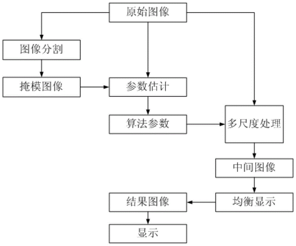
[0054] Such as figure 1 As shown, a balanced display method of a flat-panel X-ray image in this embodiment includes the following steps:

[0055] (1) Input the original image, divide the original image into several regions according to the difference of pixel gray value, and generate the mask image;

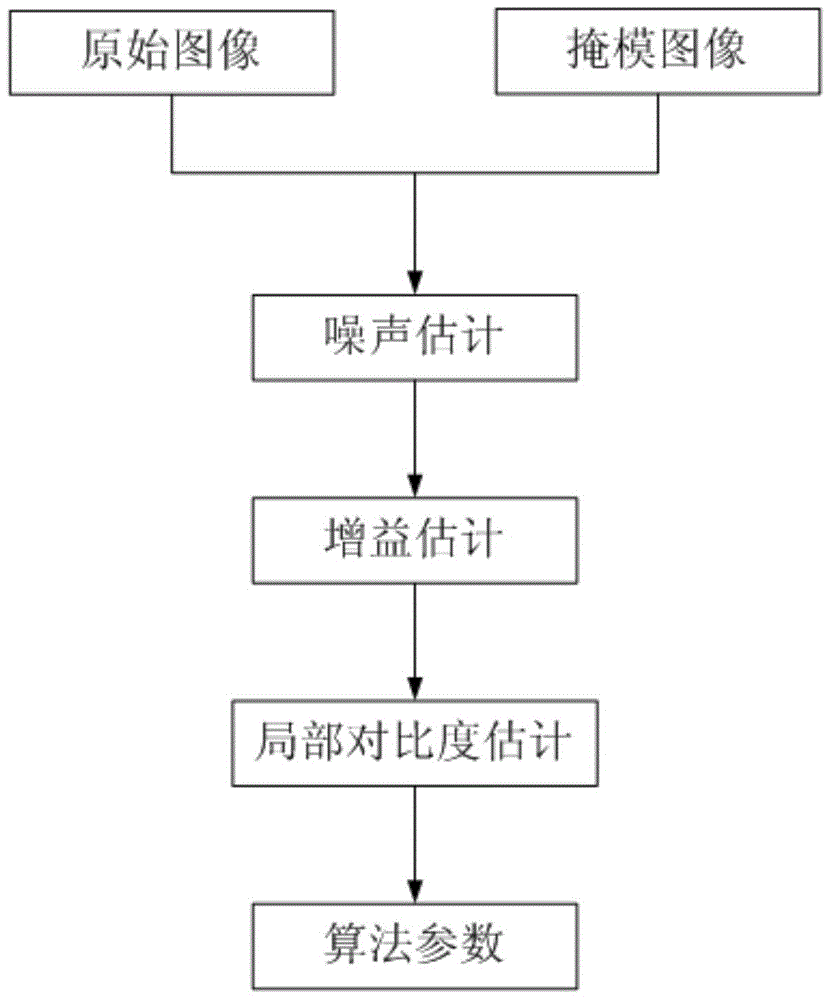
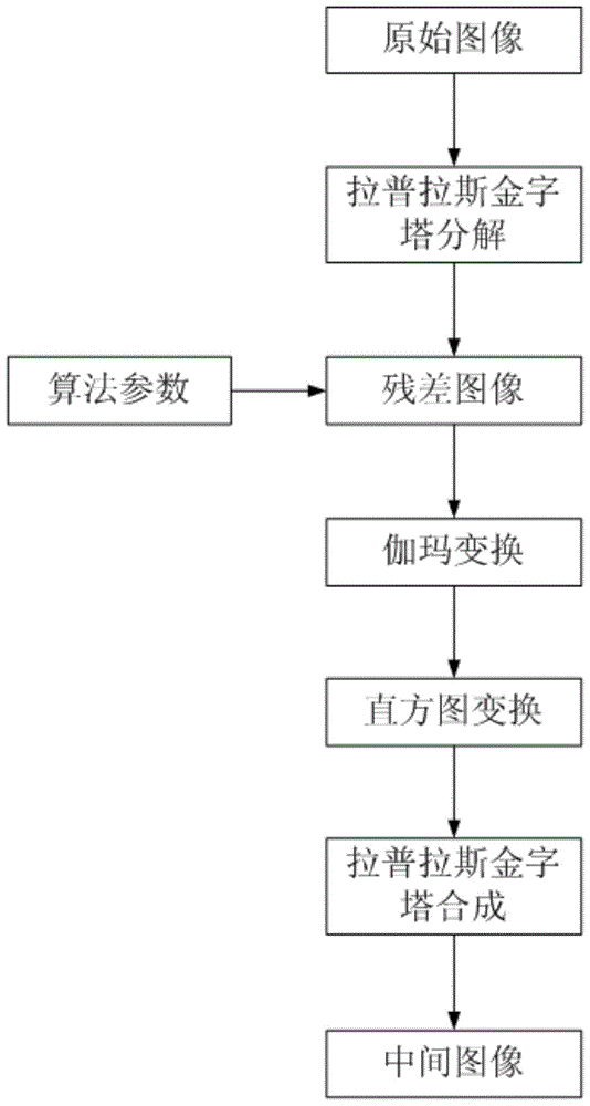
[0056] (2) Parameter estimation: read in the original image and mask image, perform noise estimation, gain estimation, and local contrast estimation in sequence, and use the estimated results as algorithm parameters; figure 2 shown, including:

[0057] (2-1) read in the original image;

[0058] (2-2) Read in the mask image;

[0059] (2-3) Noise estimation, first global noise estimation, and then perform noise estimation of each area for different areas of the mask image to generate noise parameters: Assuming the original image I, the mask image is divided into three different ones obtained by thresholding area M 1 ,M 2 and M 3 , for example, three different regions of bon...


PUM


Abstract

The invention discloses a balanced display method for a flat-plate X-ray image. The method includes the following steps of firstly, inputting an original image, segmenting the original image into a plurality of regions according to the differences of pixel point grey values so as to generate a mask image; secondly, reading in the original image and the mask image, sequentially conducting noise estimation, gain estimation and local contrast ratio estimation, and synthesizing the estimation results into algorithm parameters; thirdly, conducting Laplacian pyramid decomposition on the original image to obtain a residual error image, sequentially conducting gamma conversion and histogram conversion on the residual error image according to the algorithm parameters obtained in the second step, and then conducting Laplacian pyramid composition to generate an intermediate image; fourthly, constructing an adjustment curve, mapping the intermediate image into a result image according to the adjustment curve, calculating the optimized window width value and the optimized window position value, and displaying the result image on a display. By means of the balanced display method, target characteristics, at different scales, in the image can be effectively enhanced, and automatic optimal balanced display is achieved for the image.

Description

technical field [0001] The invention relates to digital image processing and display technology, in particular to a method and system for balanced display of flat-panel X-ray images. Background technique [0002] X-ray flat panel detectors are mainly used in medical institutions and industrial flaw detection. It is the latest and most advanced X-ray acquisition device, which can capture tiny details. The bit depth of the collected images is as high as 14 bits, which is equivalent to 16384 gray levels. Much larger than the 8 or 10 bits of image intensifiers widely used today. From the original 14-bit image data collected, to the 12-bit professional grayscale display, to the 6-bit grayscale resolution of the user's human eye, the grayscale is gradually decreasing, so the original image cannot be directly used by the end user , must undergo a series of processing such as feature enhancement, noise suppression, dynamic range compression, and contrast enhancement before it can b...

Claims


Application Information

Patent Timeline
Patent Type & Authority Patents(China)
IPC IPC(8): G06T5/00G06T5/40
Inventor 吴元黄茜黄震川
Owner 深海精密科技(深圳)有限公司
Why Eureka
Try Eureka