Contusive retina internal segment and external segment deficiency detecting method based on SD-OCT

A technology of SD-OCT and detection method, which is applied in the direction of eye testing equipment, medical science, instruments, etc., and can solve problems such as poor accuracy, large missing volume error, and unbalanced classification

Inactive Publication Date: 2014-11-12
SUZHOU UNIV
View PDF1 Cites 18 Cited by
  • Summary
  • Abstract
  • Description
  • Claims
  • Application Information

AI Technical Summary

Problems solved by technology

The research in this area is still in its infancy. The existing technology detects the absence of inner and outer retinal segments only for a small local area. The feature extraction is not comprehensive and the classification is unbalanced. For example, CN103679198A discloses a method based on K-nearest neighbors. Automatically detect the absence of inner and outer retinal segments, extract the inner and outer segment areas centered on the center of the macula and have a diameter of 1 mm as the region of interest, and perform five feature extractions. The extracted features are few, and the features are not typical enough, and because the K nearest neighbor classifier However, the K-Nearest Neighbor Classification has a classification imbalance problem, and the classification performance is poor; secondly, in the post-processing and missing volume calculation, the results obtained by the recognition classification are not excluded from the influence of vessel outline and outlier points, and the error of the missing volume is large. , poor accuracy

Method used


Image

  • Contusive retina internal segment and external segment deficiency detecting method based on SD-OCT
  • Contusive retina internal segment and external segment deficiency detecting method based on SD-OCT
  • Contusive retina internal segment and external segment deficiency detecting method based on SD-OCT

Examples


Embodiment Construction

[0071] The present invention will be further described below in conjunction with the accompanying drawings.

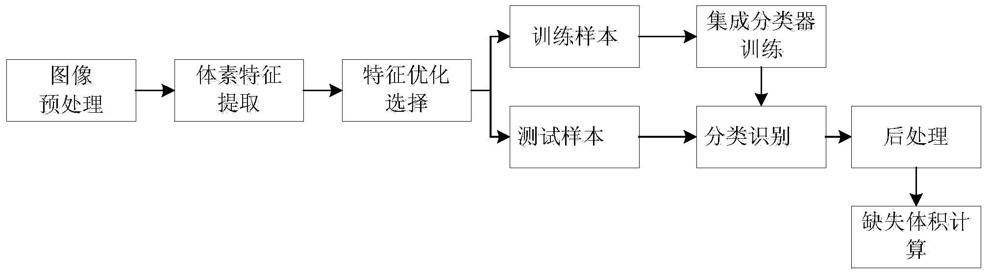
[0072] The basic block diagram of this method is attached figure 1 As shown, it mainly includes 6 steps: image preprocessing, voxel feature extraction, feature optimization selection, integrated classifier training, voxel missing / non-missing classification recognition, post-processing and missing volume calculation. The specific description is as follows.

[0073] (1) Image preprocessing

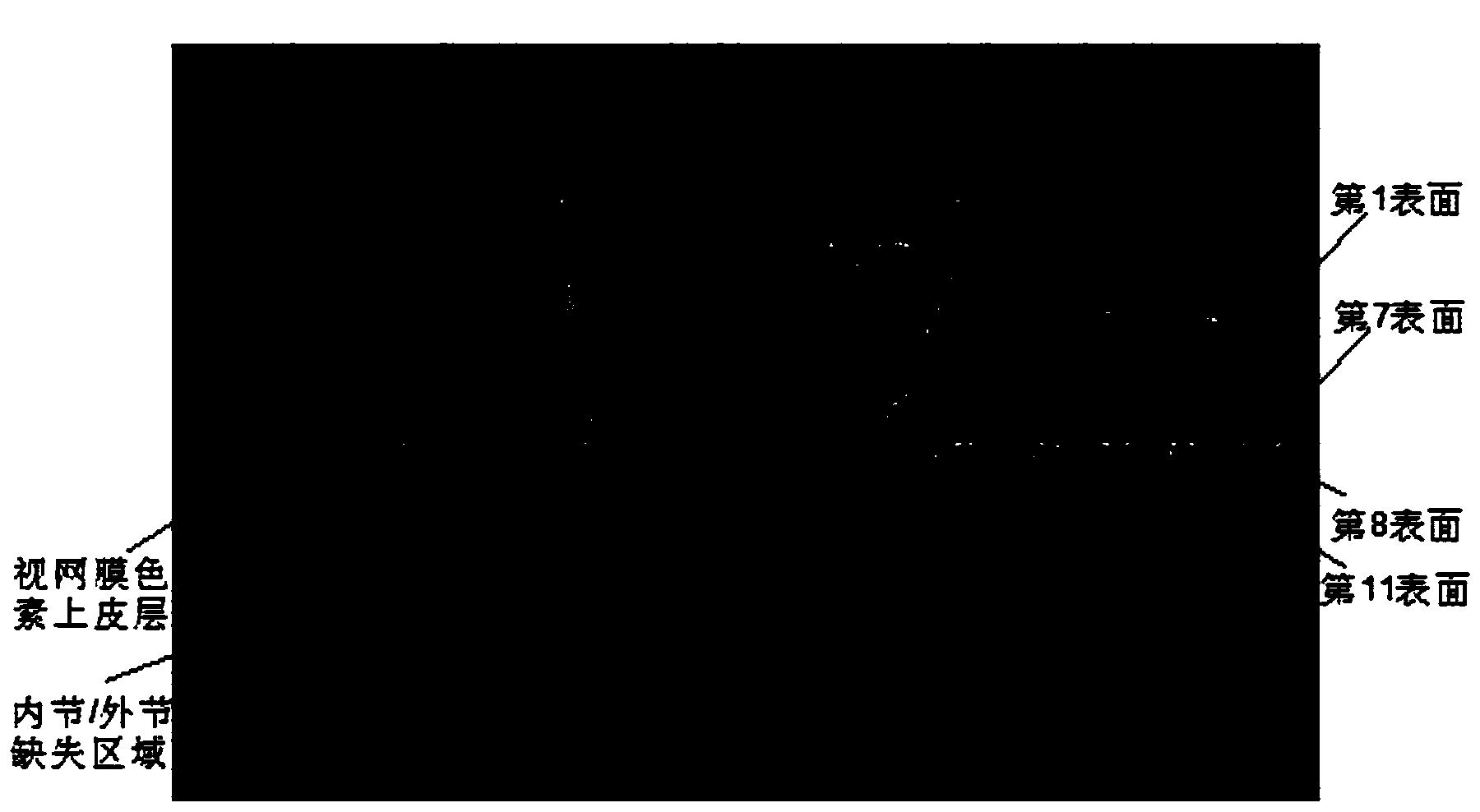
[0074] Image preprocessing mainly includes the following three steps: internal layering of the retina, extraction and flattening of the region of interest including the inner segment / outer segment region, and bilinear filter enhancement of the image.

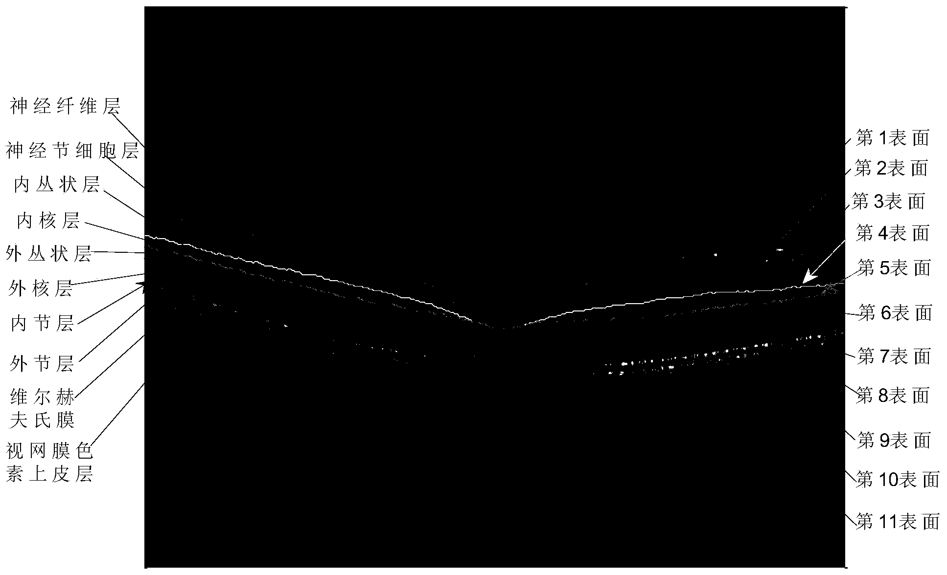
[0075] (a) Inner retinal layers

[0076] The human retina is a fairly thin tissue, less than 0.5mm thick, and is the most important component of the eye. The retina itself has a fairly complex structure, the basic structure of whic...


PUM


Abstract

The invention discloses a contusive retina internal segment / external segment deficiency three-dimensional automatic detecting method based on an SD-OCT image. The method comprises the following steps that (1), an image is preprocessed, wherein the interior of the retina is automatically divided into eleven surfaces through a multi-scale three-dimensional image division method, an internal segment / external segment area between the seventh surface and the eighth surface is extracted as an area of interest, and planarization processing and bilinear filtering enhancement are carried out; (2), five classes (fifty-seven in total) of features are extracted from each voxel in the area of interest; (3), the features are optimized and selected through a principal component analysis method; (4), the feature samples are classified to be a training set and a testing set, and the samples of the training set are trained and integrated to be a classifier through an Adaboost algorithm; (5), deficiency / non-deficiency identification is carried out on the samples of the testing set; (6), postprocessing such as vascular contour influence elimination and isolated point elimination are carried out on an identification result, the corresponding deficiency size is calculated, the identification errors of the deficiency size are small, and accuracy is good.

Description

technical field [0001] The invention belongs to retinal image processing and analysis methods, in particular to a three-dimensional automatic quantitative detection method for inner segment / outer segment loss in SD-OCT (frequency domain optical coherence tomography) retinal images. Background technique [0002] Ocular trauma is one of the main factors leading to visual impairment and even blindness, and it is the first cause of blindness in monocular blindness in my country. When damage to the surface of the eyeball propagates to the posterior retina, concussion retinal injuries are characterized by grayish-white discoloration, or when the trauma is closed, the retina remains cloudy. Histopathological studies of human eyes and animal studies have shown that damage to the inner segment / outer segment junction of photoreceptors is the etiology of retinal concussion injury. The inner / outer segments of photoreceptors are closely related to light transmission, therefore, the inte...

Claims


Application Information

Patent Timeline
Patent Type & Authority Applications(China)
IPC IPC(8): G06K9/00G06K9/36G06K9/46A61B3/10
Inventor 陈新建朱伟芳张立春陈浩宇石霏向德辉王莉芸张莉高恩婷
Owner SUZHOU UNIV
Why Eureka
Try Eureka