Root canal preparation effect detection method and device in stomatology teaching

A detection method and root canal technology, applied in the field of stomatology teaching, can solve the problems that the overall situation of the root canal cannot be accurately grasped, the isolated tooth model cannot evaluate the effect of students well, and there are no teaching models and solutions, etc. To achieve the effect of improving quality

Active Publication Date: 2015-01-21
TONGJI HOSPITAL ATTACHED TO TONGJI MEDICAL COLLEGE HUAZHONG SCI TECH
View PDF9 Cites 7 Cited by
  • Summary
  • Abstract
  • Description
  • Claims
  • Application Information

AI Technical Summary

Problems solved by technology

However, the disadvantage of this method is that whether it is split longitudinally or transversely, it can only reflect the local shape of the root canal, and cannot accurately grasp the overall situation of the root canal.
Therefore, the above two isolated tooth models cannot be used to evaluate the effect of root canal preparation and irrigation for students
[0005] At the same time, it is worth noting that in the past, teachers only relied on visual observation and experience judgment to evaluate the quality of root canal preparation of students, which could not avoid errors caused by visual deviation and different evaluation standards among different teachers, so it was difficult to guaran

Method used


Image

  • Root canal preparation effect detection method and device in stomatology teaching
  • Root canal preparation effect detection method and device in stomatology teaching
  • Root canal preparation effect detection method and device in stomatology teaching

Examples


Embodiment 1

[0044] 1) The extracted real isolated teeth were collected from the clinic. Here, the first premolar is taken as an example. The root canal system of the real isolated teeth was scanned by micro-CT. Based on the obtained original DICOM data, it was obtained by three-dimensional processing with Mimics software. Macroscopic and microscopic bionic 3D printing STL files, and STL file data for model reconstruction, submitted to high-precision 3D printers, using transparent resin materials to produce transparent resin tooth models. The root canal system of this transparent resin tooth model before root canal preparation was stained with blue dye.

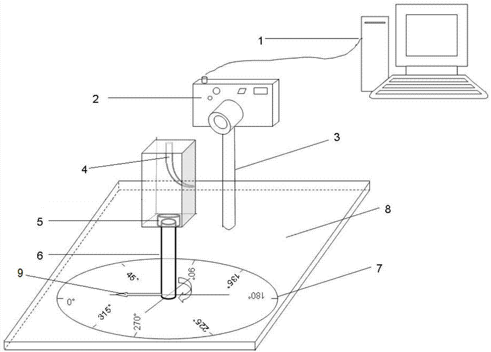
[0045] 2) Fix the transparent resin tooth model with the root canal system dyed blue on the figure 1 On the abutment 5 of the root canal preparation effect detection device in the oral medicine teaching shown.

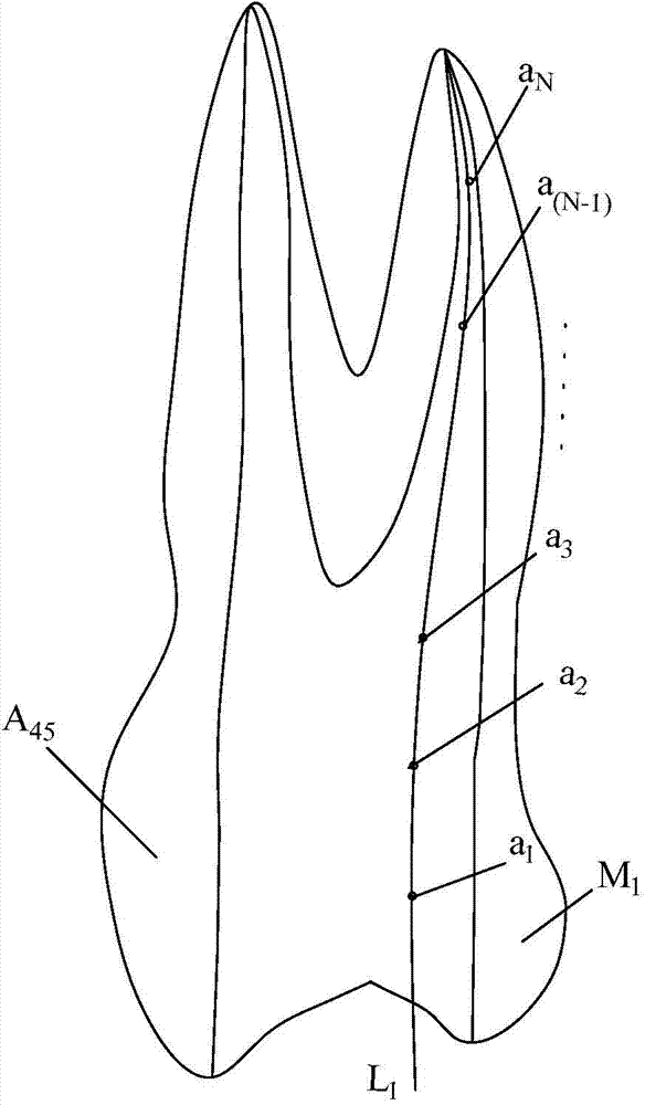
[0046] 3) Take the crown and buccal surface of the transparent resin tooth model as the frontal plane, adjust the focal length ...

Embodiment 2

[0058] 1) A commercially available transparent resin simulated root canal 4 with standardized overall length and curvature is selected, and the root canal system of the transparent resin simulated root canal 4 before root canal preparation is stained with blue dye.

[0059] 2) Fix the root canal system 4 with transparent resin dyed blue to simulate the root canal figure 1 On the abutment 5 of the root canal preparation effect detection device in the oral medicine teaching shown.

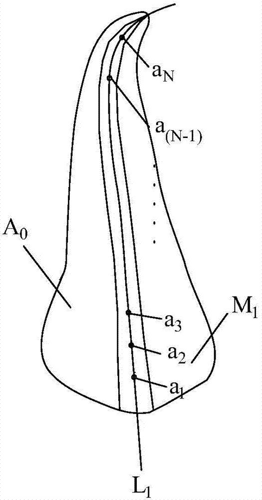
[0060] 3) Take the central axis plane that fully displays the root canal bending direction as the positive plane, adjust the focal length of the digital camera 2 to 30cm, and fix the focus of the transparent resin simulated root canal 4 in a direction perpendicular to the positive plane with the lens of the digital camera 2 Shooting, now the pointer 9 points to the position where the degree on the protractor 7 is 0°, and the frontal image A before the preparation is obtained 0 ,Such as figure 2 sh...


PUM

PropertyMeasurementUnit
Focal lengthaaaaaaaaaa

Abstract

The invention discloses a root canal preparation effect detection method and device in stomatology teaching. The method is characterized by dying a transparent resin tooth model or a transparent resin simulated root canal before and after the root canal preparation with blue dye and red dye respectively and then carrying out digital fixed focus shooting; extracting contour of the outer wall of the root canal before and after preparation and the central axis of the root canal before and after preparation in shot mages by utilizing image processing software, and setting a plurality of mark points before and after preparation at intervals on the central axis of the root canal before and after preparation; and finally, carrying out overlapping comparison on the corresponding images, determining the relative positions of the corresponding mark points on the comparison image to detect and estimate the preparation effect of the root canal. The device is mainly formed by a protractor arranged on a base, a rotation shaft arranged in the center hole of the protractor, a base platform arranged at the upper end of the rotation shaft and used for fixing the transparent resin tooth model or the transparent resin simulated root canal, and a digital camera connected with a computer. According to the root canal preparation effect detection method and device in stomatology teaching, students and teachers can visually estimate the change of shape of the root canal before and after preparation conveniently.

Description

technical field [0001] The invention belongs to the field of stomatology teaching, and specifically refers to a method and a device for detecting root canal preparation effects in stomatology teaching. Background technique [0002] Pulp disease and periapical disease are common inflammatory diseases in the oral cavity caused by various external factors. Root canal therapy is currently the most effective treatment for pulp disease and periapical disease. It achieves the purpose of completely removing inflammatory and necrotic substances in the pulp cavity and periapical tissue through perfect root canal preparation. Therefore, the success of root canal therapy depends largely on the quality of the root canal preparation. Root canal preparation includes mechanical preparation and root canal irrigation. High-quality root canal preparation should be through mechanical preparation of root canal instruments and thorough chemical irrigation to basically remove the debris and smear...

Claims


Application Information

Patent Timeline
IPC IPC(8): G09B23/28
CPCG09B23/283
Inventor 马净植王丹
Owner TONGJI HOSPITAL ATTACHED TO TONGJI MEDICAL COLLEGE HUAZHONG SCI TECH
Why Eureka
Try Eureka