Vaginal dilator with image collection function and image collection system based on vaginal dilator

A technology of image collection and vagina dilator, which is applied in the field of medical devices, can solve the problems of inconvenient use, poor observation effect, doctors' inability to diagnose patients' diseases, etc., and achieve the effect of expanding the scope of application and facilitating the comparison and summary of diseases

Active Publication Date: 2015-03-04
JIANGMEN DACHENG MEDICAL EQUIP
View PDF10 Cites 11 Cited by
  • Summary
  • Abstract
  • Description
  • Claims
  • Application Information

AI Technical Summary

Problems solved by technology

[0003] The existing vaginal dilator has the following disadvantages during use: when a doctor or nurse uses the vaginal dilator, he needs to find a light source to cooperate with it. The common situation is that he takes the vaginal dilator in one hand and the light source in the other hand, or needs to Other people cooperate, which causes inconvenience to use; in addition, some existing vaginal dilators with their own light sources only have a white light source, and some secretions or diseased substances in the vagina cannot be clearly seen under the irradiation of white light. display, it needs to be clearly displayed under blue light or green light
However, existing colposcopes have many disadvantages when in use: 1. Due to the long-distance fixed-point observation, the observation effect is poor and there are dead spots; It brings great inconvenience; 3. The colposcope ...

Method used


Image

  • Vaginal dilator with image collection function and image collection system based on vaginal dilator
  • Vaginal dilator with image collection function and image collection system based on vaginal dilator

Examples


Embodiment Construction

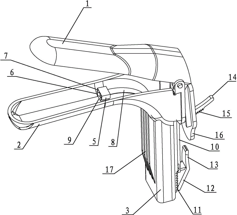
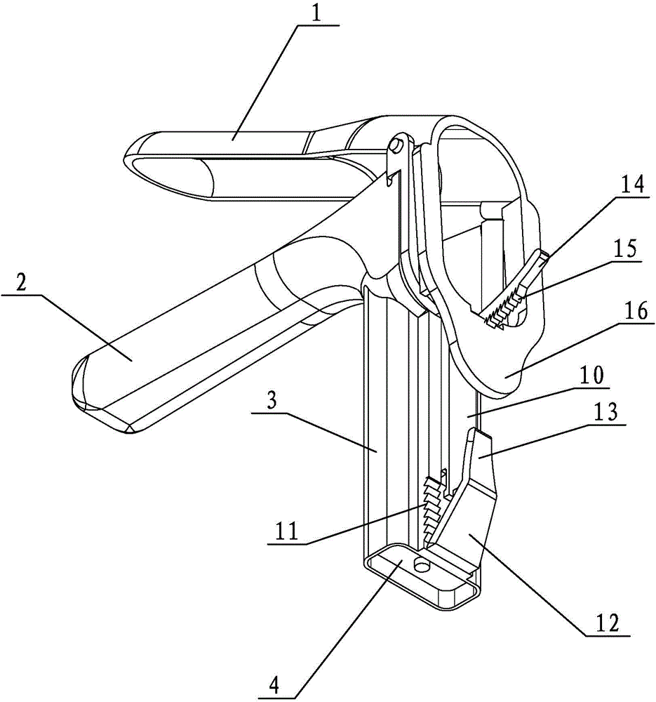
[0028] refer to figure 1 , figure 2 , the preferred embodiment provided by the present invention, a vaginal dilator with image collection function, includes an upper support 1 and a lower support 2, and a handle 3 is integrally provided under the lower support 2. The front of the handle 3 is provided with anti-slip lines 17 , and the rear portion of the handle 3 is provided with an up and down chute on which a connecting bracket 10 is slidably installed, and the upper support 1 is hinged on the top of the connecting bracket 10 . The handle 3 is provided with a cavity, and the electronic controller 4 is installed in the cavity of the handle 3 .

[0029] The two sides of the chute are provided with limited first barbs 11, and the connecting bracket 10 is provided with an inclined clamping plate 12 matched with the first barbs 11. When the connecting bracket 10 slides upwards, the clamping plate 12 is stuck on the first barbs. 11, fix the position of the connecting bracket 10....


PUM


Abstract

The invention discloses a vaginal dilator with an image collection function and an image collection system based on the vaginal dilator. The vaginal dilator comprises a lower supporting piece and an upper supporting piece. The lower supporting piece is provided with a handle. An electronic controller is arranged in the handle. A probe is arranged at the front end of the electronic controller and extends to the space between the upper supporting piece and the lower supporting piece. The probe is provided with a camera lens and at least two LED lamps different in color. The LED lamps include the white LED lamp and the blue LED lamp or the green LED lamp. The electronic controller is provided with an LED control circuit which is connected with the LED lamps, a temporary storage module connected with the camera lens, and a wireless transmission module connected with the camera lens. The electronic controller is further provided with a battery for supplying power to the modules. By means of the vaginal dilator, the situation inside a vagina can be conveniently and comprehensively observed and shot, and the vaginal dilator can be independently used and is wide in application range. In addition, the vaginal dilator can be used in cooperation with the cloud storage technology for establishing the medical vagina image collection system.

Description

technical field [0001] The invention relates to the field of medical devices, in particular to a vaginal speculum with image collection function and an image collection system based on the vaginal speculum. Background technique [0002] At present, doctors mainly rely on vaginal dilators and colposcopes to examine gynecological patients. The vaginal dilators are used to dilate the vagina. Under the irradiation of external light sources, doctors can examine the cervix. The colposcope must be used with the help of vaginal dilators. The best method of use is to first open the vagina with a vaginal dilator, place the colposcope at a certain distance outside the vagina, and observe, photograph and collect images of the patient's vagina with the help of an external light source. [0003] The existing vaginal dilator has the following disadvantages during use: when a doctor or nurse uses the vaginal dilator, he needs to find a light source to cooperate with it. The common situation...

Claims


Application Information

Patent Timeline
IPC IPC(8): A61B1/303A61B1/32A61B1/00A61M29/00
CPCA61B1/00006A61B1/00009A61B1/00016A61B1/0002A61B1/303A61B1/32
Inventor 陈志龚政龚贤应
Owner JIANGMEN DACHENG MEDICAL EQUIP
Why Eureka
Try Eureka