Method and system for adaptive image guided intervention

A technology for medical intervention and imaging data, applied in the field of medical imaging, which can solve the problems of increasing the radiation dose of medical professionals and slowing down the progress of intervention.

Inactive Publication Date: 2015-03-25
KONINKLJIJKE PHILIPS NV
View PDF5 Cites 7 Cited by
  • Summary
  • Abstract
  • Description
  • Claims
  • Application Information

AI Technical Summary

Problems solved by technology

However, repeated imaging scans during an interventional procedure undesirably increase the radiation dose delivered to the patient and / or the medical professional performing the intervention and also slow down the progress of the intervention

Method used


Image

  • Method and system for adaptive image guided intervention
  • Method and system for adaptive image guided intervention
  • Method and system for adaptive image guided intervention

Examples


Embodiment Construction

[0016] The present disclosure describes methods and systems for adaptive image-guided medical interventions using a surface attached to the patient's body at the interventional insertion site and / or near the target anatomy of interest. Chip optical shape sensing. The methods and systems may be used in conjunction with one or more imaging modalities, such as computed tomography (CT), magnetic resonance imaging (MRI), ultrasound, fluoroscopy, and the like.

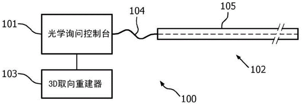
[0017] figure 1 The optical sensing system 100 is schematically illustrated. The optical sensing system 100 has an optical interrogation console 101 optically connected to at least one optical sensor device 102 and electrically connected to a three-dimensional orientation reconstructor 103 . Optical interrogation console 101 sends optical interrogation signals to optical wires 104 embedded within or on substrate material 105 to form optical sensor device 102 . The optical signal travels out to the optical sensor device 10...


PUM


Abstract

A method and system for generating imaging data during a medical intervention are provided. An external optical sensor is attached on the patient's body, and includes at least one external orientation fiber core configured to measure an orientation of the external optical sensor relative to a point of reference. The external optical sensor is interrogated to generate external sensor orientation information during the medical intervention. That information is used to estimate an orientation of the external optical sensor, which is then displayed during the medical intervention.

Description

technical field [0001] The present application relates generally to methods and systems for use in the field of medical imaging, and in particular to adaptive image-guided medical interventions. An "intervention" in this context is the insertion of one or more medical instruments into a patient in order to perform a medical procedure. Medical instruments can be such as needles, mechanical scalpels (lancets), laser scalpels, endoscopes, microscopic imaging probes, surgical staplers, retractors, cautery devices (electrical or optical), catheters, chisels , clamps, probes, trocars, scissors, etc. During many interventional procedures, a physician will use more than one medical device. Background technique [0002] Such medical interventions are often performed in conjunction with preoperative and / or intraoperative imaging scans of the patient to guide instrument manipulation during the procedure. Preoperative or scout volumetric imaging performed with modalities such as comp...

Claims


Application Information

Patent Timeline
Patent Type & Authority Applications(China)
IPC IPC(8): A61B5/00A61B5/06A61B5/113A61B6/12A61B19/00
CPCA61B5/065A61B5/1135A61B5/6833A61B6/12A61B2560/0266A61B34/20A61B2034/2061A61B5/0059A61B5/107A61B5/113A61B5/6823A61B5/6848A61B5/742A61B90/37A61B2034/2065A61B2562/0266G01L1/246A61B5/0082A61B5/7475
Inventor M·格拉斯R·陈R·曼茨克
Owner KONINKLJIJKE PHILIPS NV
Why Eureka
Try Eureka