DSA (digital subtraction angiography) cerebrovascular image auto-segmenting method based on adjacent image feature point sets

An image feature point and automatic segmentation technology, applied in the field of medical image processing, can solve problems such as robustness of algorithms, insufficient calculation speed and correctness, difficult retrospective research on historical images, and inability to meet the requirements of real-time processing, etc. Achieve the effect of reducing noise, simple operation and improving accuracy

Active Publication Date: 2015-04-08
DALIAN UNIV OF TECH
View PDF4 Cites 11 Cited by
  • Summary
  • Abstract
  • Description
  • Claims
  • Application Information

AI Technical Summary

Problems solved by technology

[0005] 1: Manual registration, the operator manually performs registration, which is inefficient, and the amount of data contained in the image data set is very large, which cannot meet the requirements of clinical real-time processing;
[0006] 2: Modular matching automatic registration technology, in which the framed registration method is based on the external fiducial point features, which can obtain high accuracy and can be used as a standard for evaluating frameless registration algorithms, but its implantable characteristics will cause patients Brings a lot of pain, and at the same time it is not easy to do retrospective research on historical images
[0007] 3: Use the algorithm based on the regular grid model to automatically generate sequence control points, optimize the process of searching for relevant points according to the characteristics of the spatial arrangement of control points, and limit the search range of control points in two-dimensional space, and then use the method based on inverse stretching The pixel mapping filling algorithm of spatial transformation is used to generate the deformed mask target image and realize subtraction, but it has defects such as robustness of the algorithm, insufficient calculation speed and correctness.

Method used


Image

  • DSA (digital subtraction angiography) cerebrovascular image auto-segmenting method based on adjacent image feature point sets
  • DSA (digital subtraction angiography) cerebrovascular image auto-segmenting method based on adjacent image feature point sets
  • DSA (digital subtraction angiography) cerebrovascular image auto-segmenting method based on adjacent image feature point sets

Examples


Embodiment Construction

[0038] In order to make the object, technical solution and advantages of the present invention clearer, the present invention will be further described in detail below in conjunction with the accompanying drawings.

[0039] like Figure 1-Figure 11 As shown, a DSA cerebrovascular image automatic segmentation method based on adjacent image feature point sets is characterized in that:

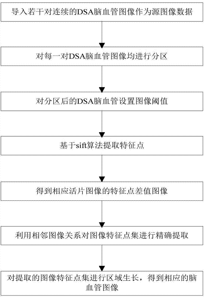
[0040] like figure 1 shown, including

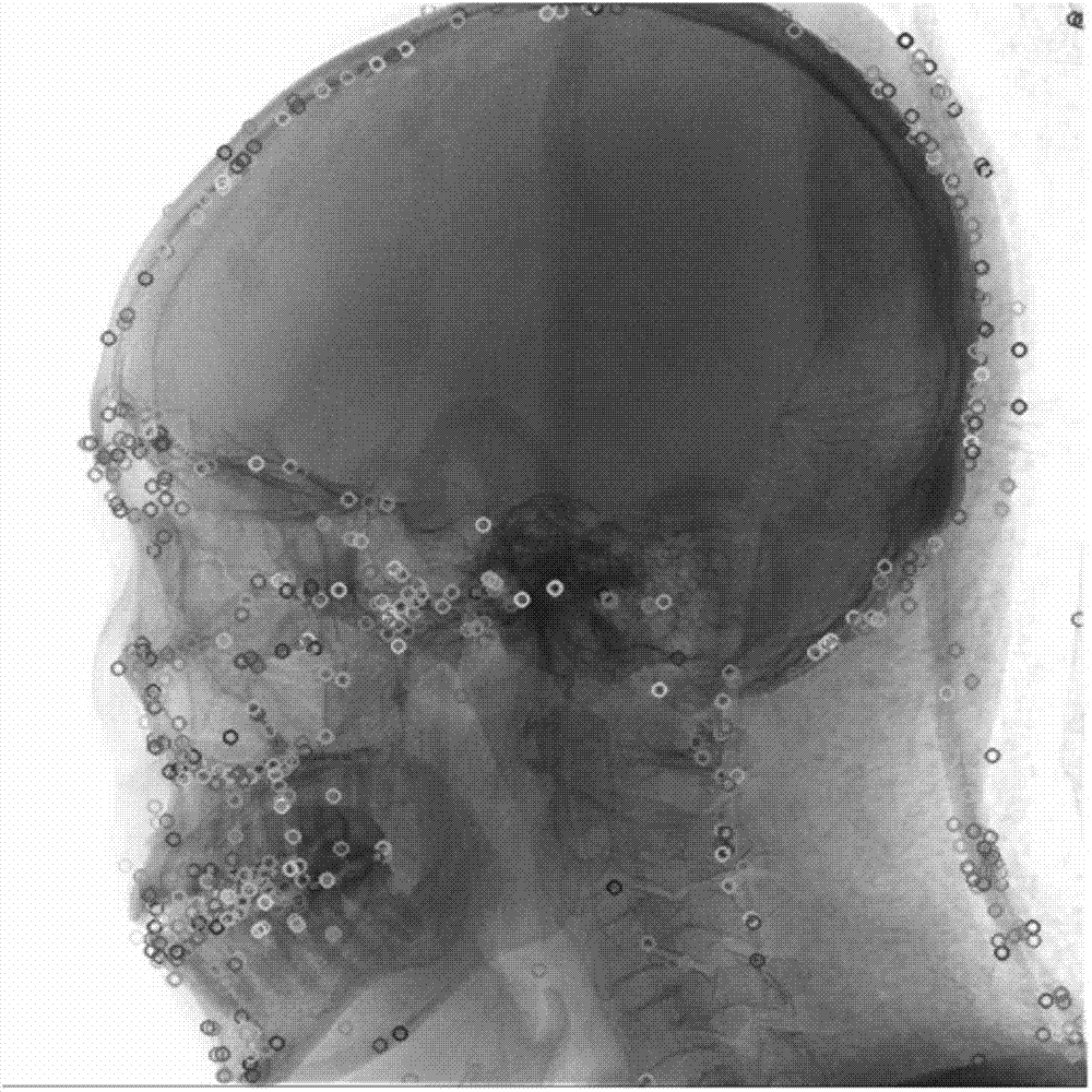
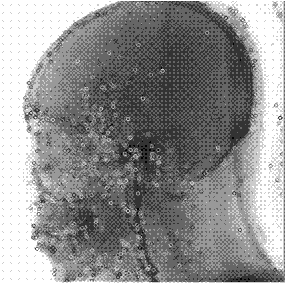
[0041] Step 1: Import several pairs of continuous DSA cerebrovascular images as source image data.

[0042] Step 2: Partition each pair of DSA cerebrovascular images, so that the mask image and live slice image in each pair of DSA cerebrovascular images are equally divided into upper and lower regions.

[0043] Further, the partition refers to all DSA cerebrovascular images (mask images and live slice images) are horizontally divided into upper and lower two regions from top to bottom from one-third of the image; The one-third part is based on experimen...


PUM


Abstract

The invention discloses a DSA (digital subtraction angiography) cerebrovascular image auto-segmenting method based on adjacent image feature point sets. The method includes: 1, importing a plurality of pairs of continuous DSA cerebrovascular images as source image data; 2, partitioning each pair of DSA cerebrovascular images; 3, setting an image threshold for each partitioned DSA cerebrovascular image; 4, extracting feature points on the basis of a sift algorithm; 5, acquiring feature point difference images of corresponding live images from mask images and live images in each pair of DSA cerebrovascular images subjected to feature point extraction, by means of the digital subtraction angiography; 6, extracting image feature point sets of all feature point difference images, and precisely extracting the image feature points by means of adjacent image relation; 7, subjecting the extracted image feature points to region growing to obtain corresponding cerebrovascular images. The method has the advantages that pixel information of adjacent domains is integrated through adjacent images by the image segmenting technique, feature point information extraction is more accurate, and noise is effectively decreased.

Description

technical field [0001] The invention relates to the field of medical image processing, in particular to a method for automatically segmenting DSA cerebrovascular images based on adjacent image feature point sets. Background technique [0002] When using DSA machine to diagnose and operate cerebrovascular malformations and cerebrovascular tumors, it usually encounters the difficulty of severe artifacts in DSA images, which seriously affects the reliability of cerebrovascular diagnosis and treatment. In order to facilitate the diagnosis of lesions, the three-dimensional information in the patient's cerebral blood vessels is obtained. The subtraction operation is usually required, and two cerebrovascular image sequences are collected, namely, the mask sequence image and the live slice image sequence; the two are correspondingly subtracted, and ideally a subtraction image containing only blood vessels should be obtained. However, the mis-matching caused by various factors often...

Claims


Application Information

Patent Timeline
Patent Type & Authority Applications(China)
IPC IPC(8): G06T7/00
CPCG06T2207/30016G06T2207/30101
Inventor 刘斌郝玲玲陈倩茹井晓彤朱琛江乾峰
Owner DALIAN UNIV OF TECH
Why Eureka
Try Eureka