Separation culture method for pig intestinal stem cells

A separation method, technology of porcine intestinal tract, applied in the field of cell separation, can solve the problem of lack of effective antibody porcine intestinal stem cell culture system

Active Publication Date: 2015-06-24
SOUTH CHINA AGRI UNIV
View PDF4 Cites 4 Cited by
  • Summary
  • Abstract
  • Description
  • Claims
  • Application Information

AI Technical Summary

Problems solved by technology

However, the isolation of intestinal stem cells from fresh pig intestines for in vitro culture is different from other species such as mice. The main difficulty lies in the lack of effective antibodies t

Method used


Image

  • Separation culture method for pig intestinal stem cells
  • Separation culture method for pig intestinal stem cells
  • Separation culture method for pig intestinal stem cells

Examples


Embodiment 1

[0031] Example 1 Isolation of porcine intestinal stem cells

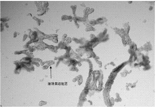
[0032] (1) Take 15-30 cm of fresh piglet jejunum, rinse the contents with HBSS, cut the intestines into 1-2 cm segments, and wash them with a separation solution (30.0 mM Na 2 EDTA-2H 2 O; 5.6mM Glucose; 5.3mM KCl; 0.45mM KH 2 PO 4 ; 4.2 mM NaHCO 3 ; 138mM NaCl; 0.34mM NaCl 2 HPO 4 ) and incubate for 30 min, remove the separation solution, resuspend these jejunum fragments with HBSS, and shake them upside down for 5-10 min to obtain porcine intestinal crypt clusters, and observe the crypt clusters under a microscope as follows: figure 1 .

[0033] (2) Add an appropriate amount of digestion solution to the obtained crypt mass, digest into single cells at 37°C, filter through a 30 μm cell sieve after termination of digestion, collect the filtrate (single cells), and resuspend these single cells with Advance DMEM-F12 , so that its concentration reaches 10 6 cells / mL.

[0034](3) First use 10% fetal bovine s...

Embodiment 2

[0036] Example 2 Two-dimensional culture of porcine intestinal stem cells

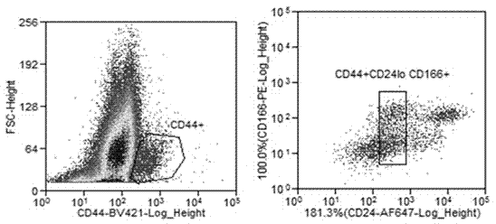
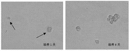
[0037] Suspend the CD44 obtained in Example 1 with complete medium containing serum and cytokines (Wnt3a, R-spondin1, Y-27632, Noggin, EGF, LY2157299 and SB202190) + CD24 lo CD166 + Cells were seeded in 96-well plates and placed in CO 2 Cultured in an incubator at a temperature of 37°C with 5% CO in the incubator 2 of humid air. By culturing, CD44 + CD24 lo CD166 + The growth of cells under two-dimensional culture conditions, the experimental results are as follows: Figure 4 .

[0038] The results showed that porcine intestinal stem cells could not form gut-like structures under two-dimensional conditions.

Embodiment 3

[0039] Example 3 Three-dimensional culture of porcine intestinal stem cells

[0040] Using CD44 containing serum, cytokines (Wnt3a, R-spondin1, Y-27632, Noggin, EGF, LY2157299 and SB202190) and Example 1 + CD24 lo CD166 + The basic mixture of cells was uniformly mixed with Matrigel, seeded into a 48-well plate, and after the mixture was solidified, a complete medium (the components of the complete medium were the same as those in Examples 1 and 2) was added to cover it. By culturing, CD44 + CD24 lo CD166 + The growth of cells under three-dimensional culture conditions, the experimental results are as follows: Figure 5 .


PUM


Abstract

The invention belongs to the technical field of cell separation and particularly discloses a separation culture method for pig intestinal stem cells. The separation culture method comprises following steps: firstly screening specific antibodies applicable to the separation of the pig intestinal stem cells, wherein the antibodies include CD24, CD44 and CD166, and the antibody CD24 is derived from ML5 monoclone; screening the pig intestinal stem cells in a targeted manner by virtue of the antibodies and a flow cytometry, establishing a three-dimensional culture system, and culturing intestine-like groups. According to the separation culture method, good materials are provided for the regeneration study of epithelia of intestinal tracts and tissue specific stem cells transplantation.

Description

technical field [0001] The invention relates to the technical field of cell separation, in particular to a method for separating and culturing porcine intestinal stem cells. Background technique [0002] The vigorous proliferation and differentiation ability of intestinal stem cells (intestinal stem cells, ISCs) plays a vital role in maintaining the integrity of the intestinal mucosal barrier structure and function and in the repair of intestinal mucosal damage. Studies have found that ISC is involved in the proliferation and differentiation of intestinal mucosa and the occurrence and development of intestinal tumors. [0003] Porcine intestinal stem cells can produce new cells to maintain the daily renewal of porcine intestinal epithelium and repair porcine intestinal epithelial damage, so it has a great effect on intestinal injury repair, intestinal health and intestinal regenerative medicine. However, porcine intestinal stem cells exist in intestinal crypts, which exist ...

Claims


Application Information

Patent Timeline
IPC IPC(8): C07K16/28C12N5/074
Inventor 黎相广王修启高春起严会超傅厚龙翟振亚陈明霞
Owner SOUTH CHINA AGRI UNIV
Why Eureka
Try Eureka