Magnetic nanoparticle magnetic resonance contrast agent and method for enhancing magnetic nanoparticle relaxivity
A magnetic nanoparticle and magnetic resonance contrast agent technology, which is applied in the fields of nanochemistry and biomedicine, can solve the problems of small size, weak magnetic moment, and decreased T2 imaging performance, and achieve simple operation, enhanced transverse relaxation rate, and high transverse The effect of relaxation rate
- Summary
- Abstract
- Description
- Claims
- Application Information
AI Technical Summary
Problems solved by technology
Method used
Image
Examples
Embodiment 1
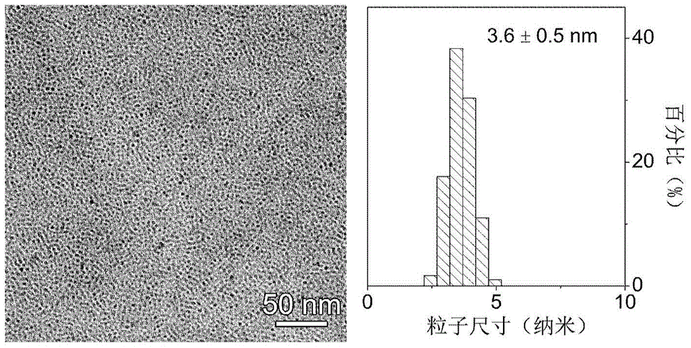
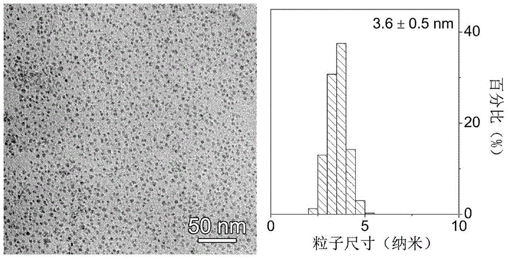
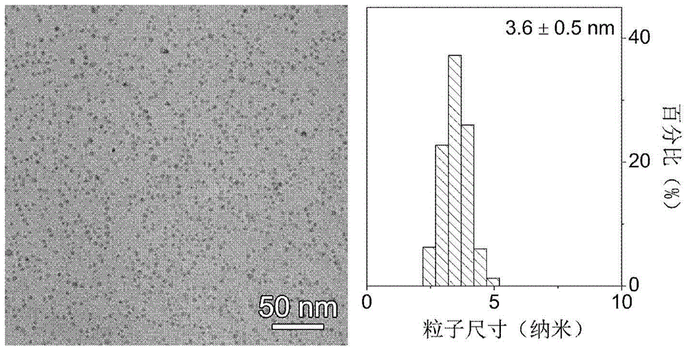
[0067] According to the literature (J.Am.Chem.Soc., 2004, 126, 273), Fe with an average particle size of 3.6 nm and a surface modified with oleic acid and oleylamine was first prepared. 3 o 4 Nanoparticles, its electron microscope photos and particle size statistics are attached figure 1 shown. Co-modify Fe with 20 mg oleic acid and oleylamine 3 o 4 Nanoparticles were dissolved in 4mL THF, and at the same time, 200 mg of polyethylene glycol 2000 (bisphosphate PEG2000, no conjugated structure in the molecule) was dissolved in 4 mL of THF, and Fe 3 o 4 The tetrahydrofuran solution and the tetrahydrofuran solution of bisphosphoric acid PEG2000 were mixed, and the ligand exchange reaction was heated at 60° C. for 8 h with stirring, and then cooled to room temperature. Add ether to precipitate, and the precipitate is magnetically separated and washed three times, and then dissolved in deionized water to obtain Fe with the surface modified by bisphosphonic acid PEG2000. 3 o 4...
Embodiment 2
[0069] According to the literature (J.Am.Chem.Soc., 2004, 126, 273), Fe with an average particle size of 3.6 nm and a surface modified with oleic acid and oleylamine was first prepared. 3 o 4 nanoparticles. Co-modify Fe with 20 mg oleic acid and oleylamine 3 o 4 Nanoparticles were dissolved in 4mL THF, and at the same time, 200mg of hydroxamic acid group-modified polyethylene glycol 2000 (hydroxamic acid PEG2000, containing p-π conjugated structure in the molecule) was dissolved in 4mL THF, and Fe 3 o 4 The tetrahydrofuran solution of hydroxamic acid PEG2000 and the tetrahydrofuran solution of hydroxamic acid PEG2000 were mixed, and the ligand exchange reaction was heated at 60° C. for 8 h with stirring, and then cooled to room temperature. Add diethyl ether to precipitate, the precipitate is magnetically separated and washed three times, and then dissolved in deionized water to obtain Fe with the surface modified by hydroxamic acid PEG2000 3 o 4 Nanoparticle magnetic re...
Embodiment 3
[0071] The 3.6nm Fe modified by bisphosphonate PEG2000 in Example 1 3 o 4 Nanoparticles and the 3.6nm Fe modified by hydroxamic acid PEG2000 in Example 2 3 o 4 Nanoparticles configured in a range of different concentrations of Fe 3 o 4 Nanoparticle solution, under the magnetic field strength of 3T, test its actual T2 MRI enhancement effect, and its T2 weighted image is as attached Figure 4 As shown, at the same Fe concentration, the hydroxamic acid PEG2000 modified Fe 3 o 4 Nanoparticles have higher contrast in T2-weighted images.
PUM
| Property | Measurement | Unit |
|---|---|---|
| particle size | aaaaa | aaaaa |
| size | aaaaa | aaaaa |
| particle diameter | aaaaa | aaaaa |
Abstract
Description
Claims
Application Information
 - R&D
- Intellectual Property
- Life Sciences
- Materials
- Tech Scout
- Unparalleled Data Quality
- Higher Quality Content
- 60% Fewer Hallucinations
Browse by: Latest US Patents, China's latest patents, Technical Efficacy Thesaurus, Application Domain, Technology Topic, Popular Technical Reports.
© 2025 PatSnap. All rights reserved.Legal|Privacy policy|Modern Slavery Act Transparency Statement|Sitemap|About US| Contact US: help@patsnap.com


