

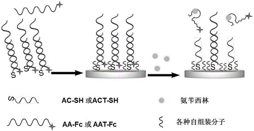
Forming method of ampicillin and sulfadimethoxine electrochemical sensor self-assembled passivation layer, and electrochemical sensor thereof
A technology of sulfadisoxine and ampicillin, applied in the field of biological analysis, achieves the effects of sensitive detection, simple design and strong versatility
- Summary
- Abstract
- Description
- Claims
- Application Information
AI Technical Summary
Problems solved by technology
Method used
Image

Examples
Embodiment 1
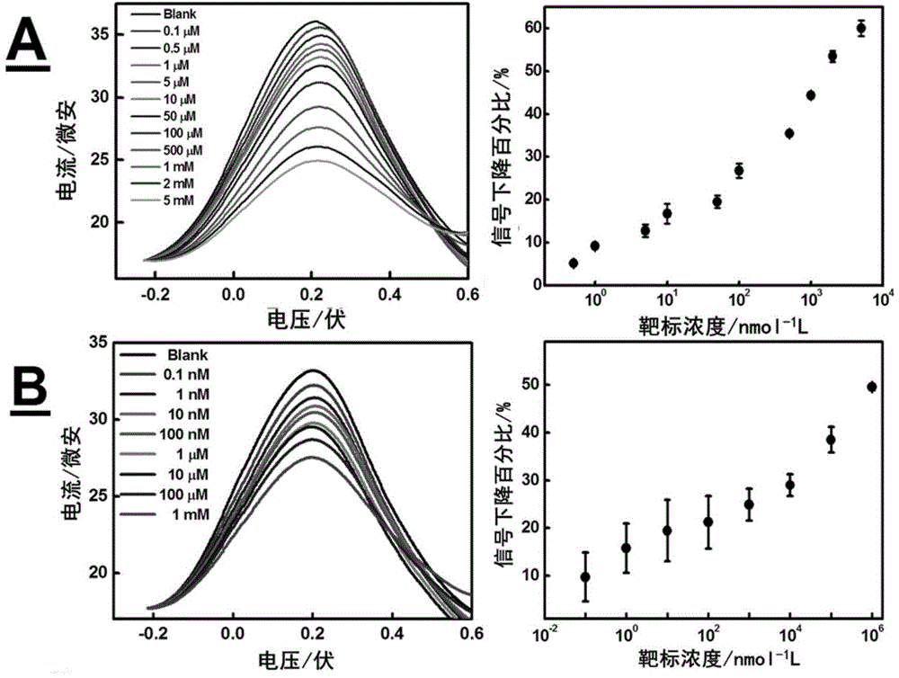
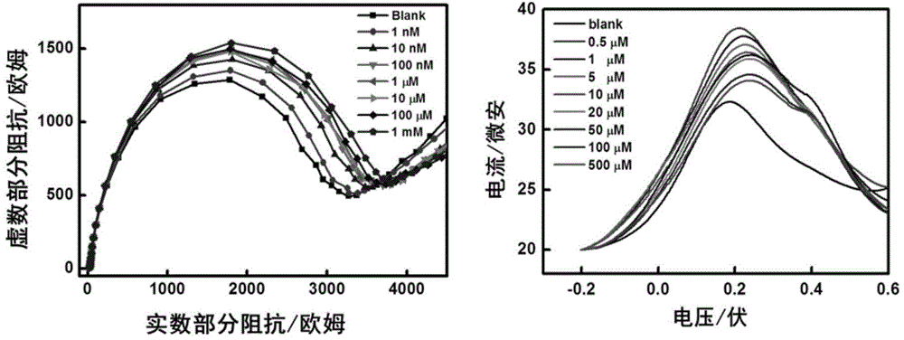
[0056]Embodiment 1: be used for the preparation of the SD-EAB A that ampicillin detects, self-assembly passivation layer is respectively OEG 6 -OMe.
[0057] Rinse the gold disc electrode (2 mm in diameter) with ultrapure water, and wash it with 1 μm, 0.3 μm, 0.05 μm Al 2 o 3 Polish the surface of the electrode with polishing powder (add a small amount of ultrapure water and solid powder on the polishing cloth and grind for 5-10 minutes), rinse with ultrapure water after each polishing, and ultrasonically in ultrapure water for 5 minutes before proceeding to the next grinding step. Ground smooth electrodes on a multi-channel potentiometer at 0.5M H 2 SO 4 The range of -0.4 ~ 1.2V is 100mV / s for 36 cycles of cyclic voltammetry scanning, the saturated mercurous sulfate electrode is used as the reference electrode, and the platinum electrode is used as the counter electrode until the cyclic voltammogram is basically stable. If no obvious corresponding redox peak is observed,...
Embodiment 2
[0059] Embodiment 2: the preparation of the sensor that is used for ampicillin detection, self-assembly passivation layer is respectively HS-(CH 2 ) 11 -(OCH 2 CH 2 ) 6 -COOH or MCH.
[0060] The self-assembled molecule in embodiment 1 is by OEG 6 -OMe was replaced by 1mM MCH or 1mMHS-(CH 2 ) 11 -(OCH 2 CH 2 ) 6 -COOH. Other steps are the same.
Embodiment 3
[0061] Embodiment 3: be used for the preparation of the SD-EAB B that ampicillin detects, self-assembly passivation layer is respectively OEG 6 -OMe.
[0062] Rinse the gold disc electrode (2 mm in diameter) with ultrapure water, and wash it with 1 μm, 0.3 μm, 0.05 μm Al 2 o 3 Polish the surface of the electrode with polishing powder (add a small amount of ultrapure water and solid powder on the polishing cloth and grind for 5-10 minutes), rinse with ultrapure water after each polishing, and ultrasonically in ultrapure water for 5 minutes before proceeding to the next grinding step. Ground smooth electrodes on a multi-channel potentiometer at 0.5M H 2 SO 4 The range of -0.4 ~ 1.2V is 100mV / s for 36 cycles of cyclic voltammetry scanning, the saturated mercurous sulfate electrode is used as the reference electrode, and the platinum electrode is used as the counter electrode until the cyclic voltammogram is basically stable. If no obvious corresponding redox peak is observed...
PUM
 Abstract
Description
Claims
Application Information